Microscopy & microtechniques
What Are the Latest Developments in Microscopy and Imaging?
Oct 28 2021
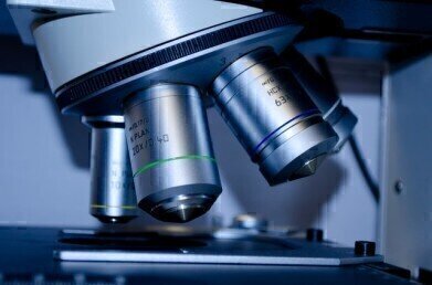
Microscopy and imaging is one of the most valuable analytical techniques available to scientists and researchers. Using state-of-the-art equipment, it’s possible to go beyond the capabilities of the naked eye and unlock incredible insight into the microscopic world. The ability to analyse cell structure and visualise biological processes at the microscopic level has contributed enormously to breakthroughs in modern science.
The industry is continually evolving, with some of the latest developments explored below:
Time-resolved electron microscopy
Advances in time-resolved electron microscopy have drastically improved biological sample imaging. The Rosalind Franklin Institute in Oxfordshire is a global leader in time-resolved electron microscopy, which uses short electron pulses to generate high-resolution images.
Scanning helium microscopy (SHeM)
Gentle and non-destructive, scanning helium microscopy uses low energy neutral helium atoms to generate detailed images of a sample’s surface. The quantum gas jet-based helium atom microscope (qHAM) is a new addition to the field and uses wave-matter duality and matter wave interference.
Introducing real-time RAPID
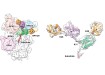
A recent paper published in the journal Nature Methods and titled ‘Universal autofocus for quantitative volumetric microscopy of whole mouse brains’ introduced the world to an exciting new analysis technique. Known as RAPID (rapid autofocusing via pupil-split image phase detection), the real-time method uses light-sheet microscopy to generate ultra-detailed images of mouse brains. This allowed the team to analyse the brain at the subcellular level, characterise 3D somatostatin-positive neurons and analyse microglia across the entire organ.
Live-cell imaging
Dutch-based company CytoSMART Technologies recently revolutionised the market with the launch of an advanced brightfield live-cell imaging system. Known as CytoSMART Lux3 BR, the microscope is equipped with a powerful 6.4 MP CMOS camera and operates within a standard cell culture incubator. It doesn’t impact temperature, airflow or culture conditions, allowing researchers to generate reliable and accurate results from live-cell imaging experiments.
“We are very excited to expand our label-free microscopy solutions for live-cell studies. For cell biologists who want to incorporate live-cell imaging into their workflow, the resolution of live-cell imaging microscopes may be too low for accurately quantifying complex read-outs, such as cell tracking or differentiation processes,” says CytoSMART Technologies CTO, Jan-Willem van Bree.
“This causes inaccurate and variable results that compromise the research output. Unlike the high-end microscopes with incubation box, the Lux3 BR ensures that cultures are not exposed to temperature, humidity, or CO2‚ fluctuations that can stress the cells, as the device easily fits in every CO2 incubator. It is a powerful addition to each research lab.”
Find out more about the game-changing new technology in ‘Label-free Microscopy Solutions for Live-cell Studies’.
.jpg)
-(2).jpg)

Digital Edition
Lab Asia Dec 2025
December 2025
Chromatography Articles- Cutting-edge sample preparation tools help laboratories to stay ahead of the curveMass Spectrometry & Spectroscopy Articles- Unlocking the complexity of metabolomics: Pushi...
View all digital editions


Events
Jan 21 2026 Tokyo, Japan
Jan 28 2026 Tokyo, Japan
Jan 29 2026 New Delhi, India
Feb 07 2026 Boston, MA, USA
Asia Pharma Expo/Asia Lab Expo
Feb 12 2026 Dhaka, Bangladesh


