-

-
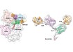
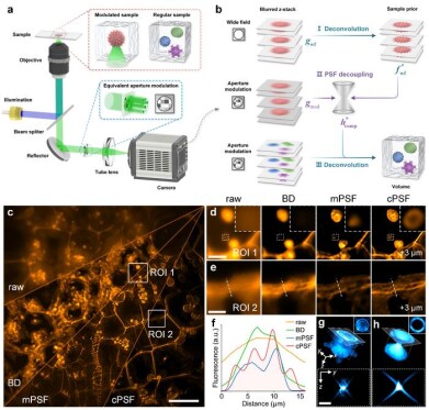 Point spread function decoupling and deconvolution. (a) Schematic of computational fluorescence microscopy. (b) Workflow of point spread function decoupling, using a fluorescent sample as a modulator and its wide-field deconvolution as a sample prior to optimise the system point spread function for subsequent deconvolution. (c) Maximum-intensity projection of a z-stack from a potato tuber sample. (d, e) Enlarged x–y views at selected regions of interest comparing raw and deconvolved images using blind deconvolution, measured point spread function and computed point spread function. (f) Cross-sectional intensity profiles from regions marked in (e). (g, h) Three-dimensional profiles with central y–z and marginal x–y slices for measured and computed point spread functions. Scale bars: 100 μm (c), 20 μm (d), 15 μm (e, g, h). Credit: Zewei Cai et al.
Fluorescence
A sample-guided approach enhances accuracy in computational fluorescence microscopy
Jan 09 2026
Researchers have reported a sample-prior-based approach to point spread function decoupling that improves system characterisation in computational fluorescence microscopy, delivering depth-extended, multicolour imaging without reliance on sub-diffraction probes or fragile theoretical models
Fluorescence microscopy remains a cornerstone of modern biological research, with widespread use to reveal cellular architecture, molecular interactions and dynamic biological processes. Computational fluorescence microscopy has extended these capabilities by combining molecular specificity with optical modulation and algorithmic demodulation, which has enabled high-resolution, multidimensional imaging beyond the limits of conventional wide-field techniques. Despite these strengths, the field has continued to face a persistent obstacle which is the accurate and practical characterisation of the imaging system itself.
Conventional strategies have tended to rely either on theoretical modelling, which rarely captures the full complexity of real optical paths and modulation schemes, or on microsphere-based measurements, which often suffer from low signal-to-noise ratios and limited depth penetration. These shortcomings have reduced imaging fidelity and have constrained the adaptability of computational fluorescence microscopy in real-world biological applications.
A team of researchers has now reported a method designed to address this long-standing problem. In a recent paper scientists, led by Professor Xiaoli Liu at Shenzhen University, China, have described a sample-prior-based point spread function decoupling strategy for computational fluorescence microscopy. The approach integrates optical modulation with computational demodulation to enable accurate system characterisation without the need to use sub-diffraction particles or to rely on theoretical assumptions.
Rather than introducing artificial probes, the method uses regular biological samples as optical modulators. These samples allow the system point spread function to be optimised computationally through a non-parametric and adaptive process. By incorporating the wide-field deconvolution result of the sample as a computational prior, the system point spread function can be refined with high accuracy while avoiding low signal-to-noise measurements and rigid parametric models.
The researchers explained that this strategy captures both system specificity and sample specificity. As a result, it supports faithful recovery of object structures even under complex optical conditions, where conventional calibration approaches often fail.
The work has demonstrated that the method can enhance computational fluorescence microscopy to achieve volumetric imaging performance comparable to confocal microscopy, alongside multicolour and depth-extended reconstruction across a range of biological tissues.
“Compared with blind deconvolution, which struggled with ill-posed optimisation, the strong support of sample priors guaranteed accurate point spread function decoupling.
“Our method not only restored fine structures such as multilayer vessels and pollen grains with high contrast but also enabled depth-extended and multichannel imaging comparable to confocal microscopy,” the authors reported.
“Using a regular sample modulator for point spread function decoupling overcame the issues of low signal-to-noise ratio and application limitation associated with sub-diffraction-limited particles, expanding the selection range and diversity of sample references for system characterisation,” they added.
The researchers also sought to highlight broader relevance for the framework.
“Although the computational fluorescence microscopy used for experimental demonstration was diffraction-limited, the proposed framework of point spread function decoupling provided a general strategy to achieve accurate system characterisation across diversified imaging modalities.
“In future research, we will design more advanced computational imaging strategies to relax the dependency on sample priors, which will enable flexible adaptation to super-resolution and dynamic live-cell imaging.
“Ultimately, this work provided a promising mechanism and method for system characterisation and demodulation to support multi-dimensional manipulation and high-performance breakthroughs in computational fluorescence microscopy,” they concluded.
Together, the findings suggest a practical route to improve robustness and performance in computational fluorescence microscopy, with implications for a wide range of biological imaging applications where accurate system calibration has remained a limiting factor.
For further reading please visit: 10.1038/s41377-025-02112-5
-(3).jpg)
Digital Edition
Lab Asia Dec 2025
December 2025
Chromatography Articles- Cutting-edge sample preparation tools help laboratories to stay ahead of the curveMass Spectrometry & Spectroscopy Articles- Unlocking the complexity of metabolomics: Pushi...
View all digital editions


Events
Jan 21 2026 Tokyo, Japan
Jan 28 2026 Tokyo, Japan
Jan 29 2026 New Delhi, India
Feb 07 2026 Boston, MA, USA
Asia Pharma Expo/Asia Lab Expo
Feb 12 2026 Dhaka, Bangladesh


