Raman spectroscopy
A novel compact Raman platform has demonstrated the ability to distinguish tumour tissue from healthy tissue with markedly improved sensitivity, raising the prospect of earlier cancer detection and wider clinical use beyond specialist laboratory settings
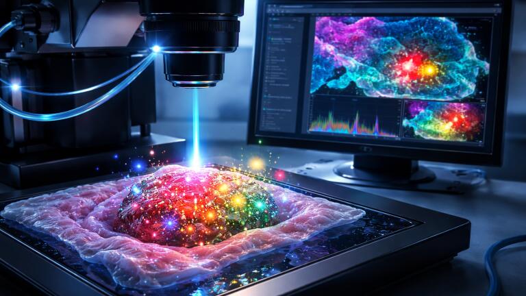
Researchers have reported the development of a novel compact Raman imaging system that has proven sensitive enough to differentiate between tumour and normal tissue. The work indicates a practical route to earlier cancer detection and to the wider adoption of molecular imaging in clinical and point-of-care settings.
The Raman system was designed to detect extremely faint optical signals emitted by surface-enhanced Raman scattering nanoparticles that bind selectively to tumour markers. After application of these nanoparticles to a tissue sample or to the area under examination, the imaging platform read the Raman signal and automatically highlighted regions likely to contain tumour tissue.
“Traditional methods for cancer-related diagnosis are time-consuming and labour-intensive because they require the staining of tissue samples and the visual assessment of abnormalities by a pathologist,” said Assistant Professor Zhen Qiu, research team leader at the Institute for Quantitative Health Science and Engineering at Michigan State University, East Lansing, Michigan.
“While our system would not immediately replace pathology, it could serve as a rapid screening tool to [aid in and] accelerate diagnosis,” he added.
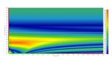
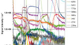
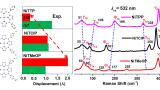
The imaging platform developed by Qiu and colleagues is able to distinguish cancerous from healthy cells by detecting Raman signals around four times weaker than those measurable with a comparable commercial system. This improvement in sensitivity was achieved through the combination of a swept-source laser – which varied its wavelength during analysis – and an ultra-sensitive detector known as a superconducting nanowire single-photon detector.
“This technology could eventually enable portable or intraoperative devices that allow clinicians to detect cancers at earlier stages, improve the accuracy of biopsy sampling and monitor disease progression through less invasive testing.
“Ultimately, such advances could enhance patient outcomes and reduce diagnostic delays, accelerating the path from detection to treatment,” said Qiu.
Qiu’s laboratory has focused on the application of superconducting nanowire single-photon detectors to enhance a range of optical imaging platforms. These detectors use a superconducting wire to register individual photons, which allows the capture of extremely weak optical signals at high speed and with very low noise.
Raman imaging enables the mapping of chemical composition by measuring the characteristic light-scattering fingerprints produced by different molecules. These signals are intrinsically weak but can be amplified through the use of surface-enhanced Raman scattering nanoparticles, which dramatically increase signal intensity when bound to specific molecular targets.
“Combining this advanced detector with a swept-source Raman architecture that replaced a bulky camera and collected light more efficiently resulted in a system with a detection limit well beyond that of comparable commercial systems.
“The fibre-coupled configuration and compact design also facilitate miniaturisation and future clinical translation,” said Qiu.
To evaluate performance, the team used surface-enhanced Raman scattering nanoparticles coated with hyaluronan acid – which promoted binding to CD44 – a surface protein expressed in many tumour cells. Initial experiments used simple nanoparticle solutions and demonstrated femtomolar sensitivity. Subsequent studies examined cultured breast cancer cells, mouse tumours and matched healthy tissues.
“The surface-enhanced Raman scattering signals were strongly concentrated in tumour samples, with only minimal background detected in healthy tissue,” said Qiu.
“This demonstrated both the exceptional sensitivity of the system and its ability to provide reliable contrast between tumour and healthy tissue. By adjusting or substituting the targeting molecule, this approach could be adapted for other cancer types,” he concluded.
The researchers noted that translation into clinical practice would require faster data acquisition and broader validation across additional tumour models and tissue types. Ongoing work has focused on improvements to imaging speed through the use of alternative laser sources, such as vertical-cavity surface-emitting lasers, or through the narrowing of the wavelength sweep range. The team has also planned multiplexing experiments that would use different nanoparticles to target multiple biomarkers at the same time with the aim to provide richer molecular information within a single scan.
For further reading please visit: 10.1364/OPTICA.569117


.jpg)
-(2).jpg)