-
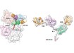
 Explainable deep learning-based unsupervised inter-domain image transformation for virtually stained high-resolution mid-infrared photoacoustic microscopy. Credit: POSTECH
Clinical
AI transforms label-free photoacoustic microscopy into confocal microscopy: a new frontier in cell imaging technology
Feb 20 2025
A team from Pohang University of Science and Technology (POSTECH), Pohang, South Korea has developed a technology that which provides stable and highly accurate cell visualisation.
In life sciences, confocal fluorescence microscopy (CFM) is highly regarded for producing high-resolution cellular images. However, it requires fluorescent staining, which poses risks of photobleaching and phototoxicity, potentially damaging the cells which are to be studied. Conversely, mid-infrared photoacoustic microscopy (MIR-PAM) allows for label-free imaging, preserving cell integrity. Yet, its reliance on longer wavelengths limits spatial resolution, making it difficult to visualise fine cellular structures with precision.
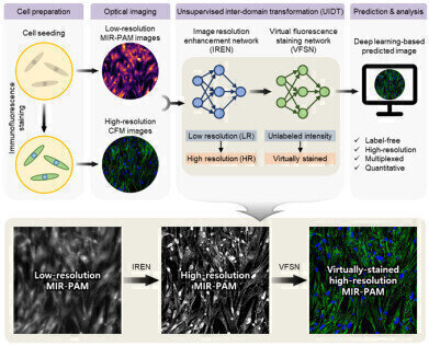
To bridge these gaps, the POSTECH team developed an innovative imaging method powered by explainable deep learning (XDL). This approach transforms low-resolution, label-free MIR-PAM images into high-resolution and virtually-stained images that resemble those which are generated by CFM. Unlike conventional AI models, XDL offers enhanced transparency by visualising the transformation process, to ensure both reliability and accuracy.
The research was led by Professor Chulhong Kim (of the departments of Electrical Engineering, Convergence IT Engineering, Mechanical Engineering, Medical Science and Engineering and the Graduate School of Artificial Intelligence) and Professor Jinah Jang (of the departments of Mechanical Engineering, Convergence IT Engineering, Medical Science and Engineering). They were assisted in their work by doctoral candidate Eunwoo Park and post-doctoral researchers, Dr. Sampa Misra and Dr. Dong Gyu Hwang.
The team implemented a single-wavelength MIR-PAM system and designed a two-phase imaging process:
- The resolution enhancement phase converts low-resolution MIR-PAM images into high-resolution ones, clearly distinguishing intricate cellular structures such as nuclei and filamentous actin.
- The virtual staining phase which produces virtually-stained images without fluorescent dyes, eliminating the risks associated with staining while maintaining CFM-quality imaging.
This innovative technology delivers high-resolution, virtually stained cellular imaging without compromising cell health, offering a powerful new tool for live-cell analysis and advanced biological research.
“We have developed a cross-domain image transformation technology that bridges the physical limitations of different imaging modalities, offering complementary benefits. The XDL approach has significantly enhanced the stability and reliability of unsupervised learning,” said Professor Chulhong Kim.
“This research unlocks new possibilities for multiplexed, high-resolution cellular imaging without labelling. It holds immense potential for applications in live-cell analysis and disease model studies,” Professor Jinah Jang added.
This research was made possible through support from the Ministry of Education, the Ministry of Science and ICT, the Korea Medical Device Development Fund, the Korean Fund for Regenerative Medicine, the Korea Institute for Advancement of Technology (KIAT), the Artificial Intelligence Graduate School Program (POSTECH), BK21 FOUR, and the Glocal University 30 Project.
For further reading please visit: 10.1038/s41467-024-55262-2
Digital Edition
Lab Asia Dec 2025
December 2025
Chromatography Articles- Cutting-edge sample preparation tools help laboratories to stay ahead of the curveMass Spectrometry & Spectroscopy Articles- Unlocking the complexity of metabolomics: Pushi...
View all digital editions


Events
Jan 21 2026 Tokyo, Japan
Jan 28 2026 Tokyo, Japan
Jan 29 2026 New Delhi, India
Feb 07 2026 Boston, MA, USA
Asia Pharma Expo/Asia Lab Expo
Feb 12 2026 Dhaka, Bangladesh


