-

-
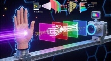 In this study, four kinds of polymer-ceramic composite scintillator films with characteristic response to different energy X-rays are prepared by a universal vitamin-assisted in-situ growth method. Through strategic architectural design of four scintillator films hetero-stacking, we achieve four-channel multi-energy X-ray curved surface imaging. This multi-energy X-ray imaging technology can effectively identify objects with different densities. Credit: Menglu Chen
X-Ray
Vitamin B1-assisted growth method enhances multi-energy X-ray imaging resolution, stability
Oct 06 2025
A Beijing Institute of Technology research team has developed a vitamin B1-assisted in situ growth method to produce perovskite polymer–ceramic scintillator films with long-term uniformity and radiation stability. The four-channel system, spanning 10 to 60 kiloelectronvolts, has achieved high-resolution imaging of both flat and curved objects, distinguishing materials such as metals and plastics in a single exposure
“Multi-energy X-ray imaging can distinguish subtle differences in material composition and density, such as skeletal abnormalities or muscular defects,” said Professor Menglu Chen of the Beijing Institute of Technology, in China.
The technology holds promise for key applications in materials science, engineering and biomedicine. However, Professor Chen noted that existing approaches have generally required a high threshold for material selection and device design.
To achieve the desired imaging resolution, it has been necessary to couple multilayer scintillators with precisely tuned energy responses, while also ensuring that each layer maintains uniformity and stability under prolonged radiation exposure. A scintillator is a material that emits visible or near-visible light when it absorbs high-energy radiation, such as X-rays, gamma rays, or energetic particles.
To address these challenges, the group has reported the development of a vitamin B1 (VmB1) assisted in situ growth method. This approach has enabled perovskite polymer–ceramic scintillator films of several different systems to achieve high levels of uniformity and radiation stability, even during long-term operation. The team emphasised that this method allows the fabrication of multilayer structures suitable for practical use in complex imaging environments.
The researchers applied density functional theory calculations to evaluate the charge distribution of polymer functional groups and their adsorption energy with perovskite. These calculations demonstrated that the stronger interaction energy between polyvinyl alcohol and the perovskite phase improved the optical performance of the scintillator films.
According to the authors, these results provided an effective theoretical framework to guide the rational selection of polymer hosts for the in-situ growth process, thereby advancing the design of robust scintillator systems.
The group further calculated the probability distribution of X-ray absorption across different energy levels within multilayer scintillator films. This work made it possible to optimise the type, thickness and stacking sequence of individual layers, thereby maximising both resolution and sensitivity. The researchers noted that the optimisation process was essential to translate the material advances into functional devices capable of broad application.
On this basis, the team successfully constructed a four-channel multi-energy X-ray imaging system covering the energy range from 10 kiloelectronvolts to 60 kiloelectronvolts. The system, realised through hetero stacking of scintillator films, was able to distinguish between different materials, such as metals and plastics, within a single exposure. This capability represents a significant advance in contrast resolution and compositional analysis, with potential to extend the diagnostic and analytical power of X-ray imaging.
In addition, the flexibility of the polymer–ceramic scintillator films has enabled high-resolution imaging of curved objects. This property has been highlighted as a major advantage for future applications, where conventional rigid detectors often fail to accommodate complex geometries.
The researchers concluded that their work has demonstrated both a material innovation and a device design strategy that together bring multi-energy X-ray imaging closer to practical deployment in fields ranging from medical diagnostics to materials technology.
For further reading please visit: 10.1186/s43074-025-00179-2
Digital Edition
Lab Asia Dec 2025
December 2025
Chromatography Articles- Cutting-edge sample preparation tools help laboratories to stay ahead of the curveMass Spectrometry & Spectroscopy Articles- Unlocking the complexity of metabolomics: Pushi...
View all digital editions


Events
Jan 21 2026 Tokyo, Japan
Jan 28 2026 Tokyo, Japan
Jan 29 2026 New Delhi, India
Feb 07 2026 Boston, MA, USA
Asia Pharma Expo/Asia Lab Expo
Feb 12 2026 Dhaka, Bangladesh


