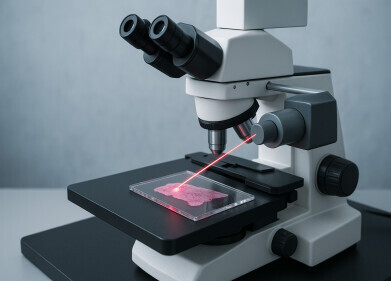
Laboratory microscope
A New Generation of Digital Cameras for Microscopy
Jun 28 2012
Motic has had only one goal in mind since 1988, and this goal is to make sure its users get products with an excellent price/performance ratio. During the decade of the 90’s its range was completed with equipment that included integrated digital cameras, and a separate range of digital cameras that could be adapted to any microscope in the market, thus offering the possibility to convert standard microscopy into digital knowledge.
In the past years there have been a great number of users that were able to upgrade their microscope with a Motic digital camera, giving the advantage of obtaining digital images in a fast and simple way. If we also take in consideration the benefits given by the images analysis software included with every model, it is plain to see that the information obtained from the captured images, allows its users to make great quality reports with reliable and precise data.
In order to keep up with the times by offering its users a modern and upgraded product, Motic launched at the beginning of 2012 a brand new range of Moticam digital cameras that has gradually substituted its former models worldwide.
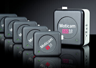
The new digital cameras with USB2.0 output are: Moticam 1 (800x600 pixels), Moticam 1SP (1,3 Mp), Moticam 2 (2,0 Mp), Moticam 3 (3,0 Mp), Moticam 5 (5,0 Mp), Moticam 10 (10,0 Mp).
To complete this new range of cameras, Motic has also added to its portfolio the Moticam 580 (5,0 Mp on SD card, HDMI output – 1080i, USB2.0 output with 800x600 pixels, Analog Video). Not only does this model allow its users to view the images by using a computer, a projector, or an HD Monitor, it also offers the possibility to capture the images directly onto an SD card at 5.0 Mp resolution, permitting its users to have more autonomy in those situations where it is not possible to have a computer at hand.
The advantages of the new Moticam range of digital cameras are:
Complete Range of CMOS cameras from 0.5 Mp until 10 Mp for the needs and the budget of every user: schools, universities, laboratories, doctors and veterinaries.
Free Software. Our cameras include the complete and actualized software Motic Images 2.0 with the possibility of live view, calibration, measurements and with several helpful tools. If you prefer to use another software, Moticam 1 and 580 are available with both Direct Show and TWAIN drivers and all the other cameras are also compatible with third party software such as Image Pro Plus 7 and Micro Manager.
Unified design. The new range of Moticam digital cameras was designed with the purpose of unifying its design with Motic’s current image.
Removable USB cable. The removable USB cable allows the user to quickly change the length of the cable, thus adjusting the distance between the camera and the computer. With this new feature the new Moticam cameras have improved its resistance by avoiding possible tensions in the USB cable.
Bigger eyepiece adapters. With the new focusable eyepiece adapters of 30 and 38mm the adaptability of the new Moticam has improved significantly.
Motic is still basing the compatibility of its new range of Moticam digital cameras in the C- Mount system, which is the most common adapter used by the majority of microscope manufacturers. This ensures the use of Moticam cameras with almost any microscope. In case it is not possible to obtain a C-Mount adapter for a certain model, Motic is still able to offer you a viable and easy alternative. All Moticam digital cameras include in its standard package a set of eyepiece adapters making it possible to easily connect the camera onto the microscope’s eyepiece.
Whether you have a biological microscope, a petrografic microscope, a metallurgical microscope or a stereo microscope, Motic is giving you the opportunity to turn your system into a digital station by converting your images into knowledge.
The software Motic Images Plus that comes with every Moticam digital camera is just as important as everything that has been mentioned previously. This software features the module MI Device that grants its users to have full control over a wide selection of parameters permitting a precise adjustment of the camera, thus enabling to capture high quality images in the best possible conditions. This software also offers the possibility to insert calibrated reticles in the image that is being viewed on the computer screen.
Once the image has been captured with the images analysis software, the user is able to make measurements and export the information to other programs (such as Excel), allowing you to use this information to make precise reports of the sample that is being analyzed.
Motic also offers optional extensions of the software, such as Motic Net which makes it possible for the user to supervise a multiple number of digital microscopy stations from a single platform (ideal for the educational field), or Motic Trace that permits its user to overlap two digital images taken from different stations in real time (ideal for forensic applications).
But this is not the end of it. At Motic we are constantly working on ways to offer our microscopy users innovative solutions at the excellent price/performance ratio that distinguishes us.
Digital Edition
Lab Asia Dec 2025
December 2025
Chromatography Articles- Cutting-edge sample preparation tools help laboratories to stay ahead of the curveMass Spectrometry & Spectroscopy Articles- Unlocking the complexity of metabolomics: Pushi...
View all digital editions
Events
Jan 21 2026 Tokyo, Japan
Jan 28 2026 Tokyo, Japan
Jan 29 2026 New Delhi, India
Feb 07 2026 Boston, MA, USA
Asia Pharma Expo/Asia Lab Expo
Feb 12 2026 Dhaka, Bangladesh
