FTIR
Spectroscopy study identifies potential biomarkers for rare metaplastic breast carcinoma tissue
Oct 08 2025
A retrospective study has provided the first comprehensive spectroscopic profile of metaplastic breast carcinoma, highlighting Fourier-transform infrared (FTIR) spectroscopy as a promising diagnostic tool to distinguish this rare tumour from ductal carcinoma in situ and invasive ductal carcinoma
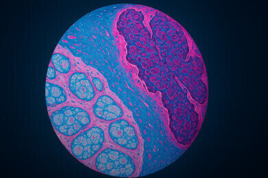
Metaplastic breast carcinoma, a rare and poorly understood form of breast cancer that accounts for fewer than 1 per cent of breast neoplasms, has now been subjected to detailed spectroscopic analysis. Researchers from the Samuel T. Adeleke' Lab at the Bowen University, Iwo, Nigeria, have reported that attenuated total reflectance Fourier-transform infrared (FTIR) spectroscopy may offer a reliable means to distinguish this malignancy from both ductal carcinoma in situ (DCIS) and invasive ductal carcinoma (IDC), as well as from normal breast tissue.
In a retrospective study, the team analysed archival tissue blocks from ten cases of metaplastic carcinoma, 12 of DCIS, and 31 of IDC. Samples were stained with haematoxylin and eosin for histological confirmation, while adjacent unstained sections were examined with FTIR spectroscopy. Tissue from ten normal breast samples served as the control group.
Statistical analyses included t-tests to determine significant differences in spectral peak intensities, hierarchical clustering to identify relationships between samples, and receiver operating characteristic (ROC) curves to evaluate diagnostic performance.
The investigators found that mean spectral peak intensities were consistently lower in all carcinoma subtypes compared with normal breast tissue. Several biochemical ratios were significantly elevated in cancerous samples, including phosphate (A1237/A1080, p < 0.01), glycogen (A1043/1543, p < 0.01), and the nucleocytoplasmic index (A1080/A1632, p < 0.03).
ROC analysis revealed that the Amide A peak at 3,280 cm−1 had an area under the curve (AUC) between 0.93 and 0.96, making it highly effective in distinguishing normal from malignant tissues. Peak 2,922 cm−1 provided moderate specificity in differentiating IDC from normal tissue (AUC ≈ 0.7), while peak 1,744 cm−1 discriminated between DCIS and metaplastic carcinoma (AUC = 0.7).
Particularly striking was the diagnostic accuracy of the 1,080/1,632 nucleocytoplasmic ratio, which achieved an AUC of approximately 1.0 in distinguishing normal from carcinoma samples. It also effectively differentiated DCIS from IDC (AUC ≈ 0.86) and DCIS from metaplastic carcinoma (AUC ≈ 0.8).
Cluster analysis reinforced these results, demonstrating biochemical signatures that linked protein, lipid, and amide peaks into consistent diagnostic patterns. Protein bands such as Amide A (3,280 cm−1), Amide I (1,632 cm−1), and β-sheet Amide II (1,543 and 1,535 cm−1) were highlighted as particularly promising biomarkers.
“Statistically significant biomarkers are important for diagnostic integration but statistical significance alone does not guarantee diagnostic utility,” the authors explained.
They emphasised that the exceptional performance of the nucleocytoplasmic ratio aligns with conventional histopathological approaches that rely on nucleocytoplasmic characterisation to assess malignancy.
The findings have provided the first comprehensive spectroscopic profile of metaplastic breast carcinoma and have underscored the diagnostic potential of FTIR spectroscopy in clinical oncology.
While the study was limited by the small number of cases available, the results have advanced understanding of the biochemical origins of this rare tumour type. The authors concluded that further studies are required to validate the approach and to develop protocols that may translate into routine biomarker use in breast cancer diagnostics.
For further reading please visit: 10.14218/ERHM.2025.00014

Digital Edition
Lab Asia Dec 2025
December 2025
Chromatography Articles- Cutting-edge sample preparation tools help laboratories to stay ahead of the curveMass Spectrometry & Spectroscopy Articles- Unlocking the complexity of metabolomics: Pushi...
View all digital editions


Events
Jan 21 2026 Tokyo, Japan
Jan 28 2026 Tokyo, Japan
Jan 29 2026 New Delhi, India
Feb 07 2026 Boston, MA, USA
Asia Pharma Expo/Asia Lab Expo
Feb 12 2026 Dhaka, Bangladesh


