-
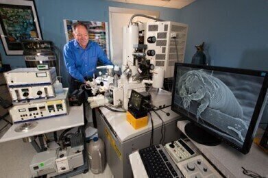
 Gary Bauchan, Director, Electron & Confocal Microscopy Unit, USDA-ARS inserts mite specimens into the Quorum PP2000 Cryo-Prep Chamber.
Microscopy & microtechniques
The Agricultural Research Service of the USDA uses a Quorum Cryo-SEM preparation system for the study of mites, ticks and other soft bodied organisms
Nov 22 2016
Quorum Technologies report on the work of the Agricultural Research Service of the US Department of Agriculture where their PP2000 Cryo-SEM preparation system is in use to prepare soft bodied organisms including mites & ticks for study using cryo-SEM
Dr Gary Bauchan is the Director of the Electron and Confocal Microscopy Unit at the Agricultural Research Service (ARS), the principal in-house research agency of the United States Department of Agriculture. The Unit is a core facility with the responsibility of providing collaborative assistance to scientists from ARS, Northeast Area and Beltsville Agricultural Research Center (BARC) who have microscopy applications that require high resolution imaging. Dr Bauchan’s team have produced images using electron microscopes of bacteria, fungi, mites, insects, nematodes and parasites along with plant and animal tissues both healthy and diseased. One of his major collaborations is with Dr Ron Ochoa, the world's expert on plant feeding mites.
Biological specimens require special treatment due to the high water content of the samples. Many of the specimens are in liquid cultures or are very soft-bodied and by using classical preparative techniques will either destroy the specimen or distort the specimen producing artefacts. A cryo-prep system is an ultra-fast method to ready the specimens for observation in a SEM especially a high resolution field emission SEM. Thus, specimens are frozen in time to allow for observation of feeding behaviour, mating behaviour, host/parasite interactions, etc. It preserves the natural orientation of ultrafine structures such as setae, antenna, legs, skin texture, sensory organs, waxy coatings and eggs.
Asked about his experience using a Quorum PP2000 Cryo-SEM preparation system on the Hitachi S-4700 field emission scanning electron microscope, Dr Bauchan said: “The Quorum system is easy to use, the set-up for imaging is logical, durable, reliable, and maintains ultra-low temperatures for a long period of time. Holders containing pre-frozen samples are transferred into the cryo-prep chamber where they are etched to remove any surface contamination (condensed water vapour) by raising the temperature of the stage from -130ºC to -90°C for 10-15 minutes. Following etching, the temperature inside the chamber was lowered below -130°C, and the specimens were coated with a 10 nm layer of platinum using a magnetron sputter head equipped with a platinum target. The specimens were transferred to a pre-cooled (-130°C) cryo-stage in the SEM for observation.”
The system has been used in multiple projects by the Unit, many of which have been published with the generation of stunning, colourful images. The use of low temperature SEM has been shown time and again to be the best method for the examination of microscopic biological specimens and their ultrastructure. The work in conjunction with Dr Ochoa has been particularly productive with five papers published this year to date. These have focused on the field of acarology, a branch of zoology dealing with the study of mites and ticks.
The PP2000 is one of Quorum's highly automated, easy-to-use, column-mounted, gas-cooled cryo-SEM preparation systems suitable for most makes and models of SEM, FE-SEM and FIB/SEM. To obtain full details of the latest cryo-SEM preparation systems and other products available from Quorum Technologies, please click here.
Digital Edition
Lab Asia Dec 2025
December 2025
Chromatography Articles- Cutting-edge sample preparation tools help laboratories to stay ahead of the curveMass Spectrometry & Spectroscopy Articles- Unlocking the complexity of metabolomics: Pushi...
View all digital editions
Events
Jan 21 2026 Tokyo, Japan
Jan 28 2026 Tokyo, Japan
Jan 29 2026 New Delhi, India
Feb 07 2026 Boston, MA, USA
Asia Pharma Expo/Asia Lab Expo
Feb 12 2026 Dhaka, Bangladesh
.jpg)
-(2).jpg)


