Microscopy & microtechniques
New Desktop Microscope Delivering Super-Resolution Performance Announced
May 25 2016
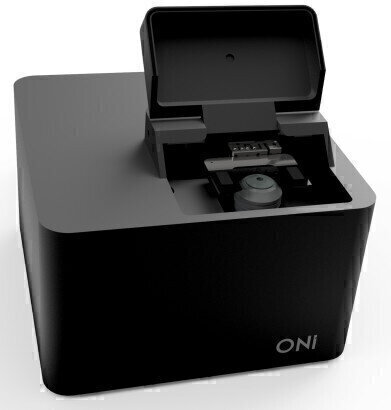
Developed in the Clarendon Laboratory in the Physics Department at the University of Oxford, Oxford Nanoimaging™ (ONI) announces the release of the exciting desktop fluorescence microscope, the Nanoimager™, to deliver nanoscale resolution imaging of live cells. This innovative approach from the Kapanidis Gene-Machines team aims to democratise the use of powerful single-molecule imaging and super-resolution microscopy technology. With a low cost of entry and ease of use, Nanoimager opens opportunities for nanoscale research without the need for a large, specialist laboratory and without a daunting training and operating burden.
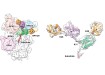
The Kapanidis group have generated stunning nanoscale images of cellular features with ten times the resolution of conventional fluorescence microscopy. Single-molecule imaging capability enables the understanding of biomolecular processes, one molecule at a time. Going further, as the most advanced commercially available FRET solution, Nanoimager can measure the dynamics of molecular interactions and structural transitions such as detecting the assembly of protein subunits or observing the synthesis of DNA in real-time.
The Nanoimager microscope unit is just 21 cm x 21 cm x 15 cm. It has been engineered from the bottom up for optimum single-molecule imaging functionality. It may be operated on a regular desk or bench without the need for extra anti-vibration or environmental isolation. The inherently robust design works together with passive dampening elements to reduce vibrations and drift, and real-time focus and sample positioning provides the stability for data collection over many hours.
The clear and intuitive user interface helps research users new to single-molecule localisation work toward rapid productivity. The large field of view, real-time data analysis features, and high degree of instrument automation enable high-throughput workflows, directly applicable to the wide range of emergent single-molecule screening applications.
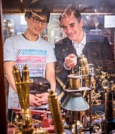
Co-inventors, Professor Achillefs Kapanidis and PhD student, Bo Jing, lead the team to break ground in this new approach to super-resolution microscopy, a project with an eight-year heritage. Professor Kapanidis said: “The new microscope can take single-molecule imaging out of physics labs and central facilities and into the hands of the chemist, the biologist, the biotechnologist. It is not only an excellent instrument for super-resolution imaging, but also a versatile, user-friendly toolbox that will help new users innovate with single molecules as their new currency.”
.jpg)
-(2).jpg)

Digital Edition
Lab Asia Dec 2025
December 2025
Chromatography Articles- Cutting-edge sample preparation tools help laboratories to stay ahead of the curveMass Spectrometry & Spectroscopy Articles- Unlocking the complexity of metabolomics: Pushi...
View all digital editions


Events
Jan 21 2026 Tokyo, Japan
Jan 28 2026 Tokyo, Japan
Jan 29 2026 New Delhi, India
Feb 07 2026 Boston, MA, USA
Asia Pharma Expo/Asia Lab Expo
Feb 12 2026 Dhaka, Bangladesh


