Microscopy & microtechniques
Innovative Stage used for the imaging of mammalian cells with Cryo-CLXM Microscopy
Nov 12 2013
Linkam Scientific Instruments report on the use of their innovative CMS196 cryo stage for the study of mammalian cells at the London Research Institute, Cancer Research UK.
In a recent publication in the journal, Ultramicroscopy (Duke et al., 2013), Dr Lucy Collinson (LRI Electron Microscopy Unit), in collaboration with Dr Sharon Tooze (LRI Secretory Pathways Lab), imaged forming autophagosomes in whole mammalian cells. The structures are particularly difficult to capture in cells prepared for electron microscopy, so they are now using a powerful new technique called cryo-soft X-ray tomography, cryo-SXT, working with Dr Liz Duke at the Diamond Light Source synchrotron. The cells are grown on tiny gold grids and plunged into liquid ethane to preserve the cells in the frozen state.
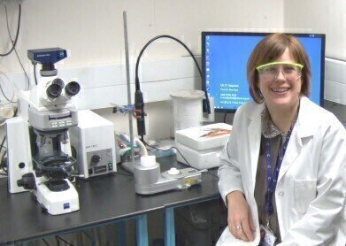
In order to find the autophagosomes within the cells, they are labelled with green fluorescent protein (GFP). The fluorescent autophagosomes are then located using a technique called correlative cryo-fluorescence and cryo-soft x-ray microscopy (cryo-CLXM). Cryo-fluorescence microscopy is performed using the Linkam CMS196 stage prior to the cells being transported in cryo-containers to synchrotrons in Oxfordshire, Berlin and Barcelona for imaging. One of the major advantages of this new correlative approach is that the CMS196 stage allows the cells to be screened for quality and protein localisation in the research laboratory before actually travelling to the synchrotron. The combination of cryo-fluorescence microscopy and cryo-SXT allows scientists to link the functionality of proteins to their near native-state structure.
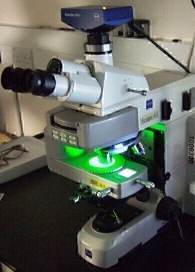
The Linkam CMS196 stage was designed specifically to solve the problem of how to get vitrified EM grids from the fluorescence microscope into the cryo-TEM without devitrification and contamination through condensation. The stage has been optimised optically to enable the use of high NA lenses. Up to 3 grids can be loaded into a specially designed cassette for transportation from the plunge freezer to the upright fluorescent microscope. The cassette is then easily loaded onto the viewing bridge using special manipulation tools. The sample viewing chamber is perfectly dry and below -180ºC while the sample bridge itself is at -196°C. The grids can be quickly and efficiently scanned using a 100X 0.75NA lens and manipulated using high precision micrometers. The cassette is then simply manipulated back into the transportation device and is then transported to the cryo-TEM under liquid nitrogen.
.jpg)
-(2).jpg)
Digital Edition
Lab Asia Dec 2025
December 2025
Chromatography Articles- Cutting-edge sample preparation tools help laboratories to stay ahead of the curveMass Spectrometry & Spectroscopy Articles- Unlocking the complexity of metabolomics: Pushi...
View all digital editions


Events
Jan 21 2026 Tokyo, Japan
Jan 28 2026 Tokyo, Japan
Jan 29 2026 New Delhi, India
Feb 07 2026 Boston, MA, USA
Asia Pharma Expo/Asia Lab Expo
Feb 12 2026 Dhaka, Bangladesh


