Laboratory events news
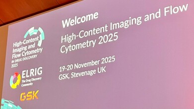
ELRIG 2025: GSK imaging strategy make cellular heterogeneity core to its drug discovery
Dec 29 2025
A dedicated bioimaging function at GSK has shown how integrated microscopy and analytics can deliver quantitative evidence across drug discovery, with a particular focus on complex therapeutic modalities and biological variability
Dr Carla Newman, associate director, cellular imaging and dynamics at GSK, outlined how a dedicated bioimaging capability at the pharmaceutical giant has supported drug discovery by aligning imaging and analytical methods closely with specific biological questions and by treating cellular heterogeneity as a core feature of biology rather than an experimental inconvenience. She described an approach that prioritised measurement, decision support and the reduction of uncertainty across discovery programmes.
Her team sits within a wider imaging capability at GSK that spans cellular microscopy through to in vivo imaging and complementary chemical characterisation for drug formulation, with substantial activity shared across both the UK and US. Within this landscape, her own group focuses on cellular imaging and dynamics, deploying both established techniques such as confocal microscopy and more specialised platforms selected to address defined challenges in modern discovery portfolios.
Her framing followed the familiar logic of drug discovery whereby a candidate must reach its site of action, engage its target, generate a functional response and do so in the correct tissue and cell type. Imaging can contribute evidence at each stage, from localisation and target expression through to quantitative measurements that feed into pharmacology, modelling and translational decision making. The aim is to deliver robust measurements that inform choices and increase the likelihood of clinical success she stressed.
A recurring theme was the shift in therapeutic modality. Small molecules remain central and benefit from decades of accumulated knowledge and predictive models. However, many current programmes involve complex modalities such as monoclonal antibodies, antibody–drug conjugates and oligonucleotide therapeutics. These introduce additional biological constraints and distinct failure modes which require adapted experimental models and measurement strategies to capture uptake, intracellular trafficking, target accessibility and variability between cells and donors.
She illustrated routine support work using innovative three-dimensional cell systems. In one programme, the question was whether organoids expressed a receptor of interest. Fluorescence labelling enabled visualisation of receptor distribution within intact organoids, combined with actin and nuclear staining and three-dimensional rendering. This allowed comparison across multiple organoids and – crucially – quantitative assessment of receptor abundance rather than reliance on qualitative judgement.
Quantification also underpinned work on antibody–drug conjugates. Pharmacokinetics and pharmacodynamics colleagues had requested metrics to strengthen patient dosing predictions. One metric concerned internalisation kinetics. Dr Newman described a pulse–chase style experiment in which cells were cooled to permit binding while suppressing uptake, exposed to a fluorescently labelled conjugate, washed and then monitored over time. Measuring intracellular fluorescence across time and concentration generated kinetic curves and parameters such as half-life that could enter modelling workflows. The value lay in producing comparable numerical descriptors across constructs.
To move beyond two-dimensional cultures, she highlighted volumetric imaging in complex biological models. Using lattice light-sheet microscopy, the team imaged organoids and air–liquid interface cultures, where ciliated epithelia form layered architectures that challenge thin optical sectioning. This enabled quantitative characterisation of donor-to-donor variation before intervention, including tissue thickness, cilia number and length, epithelial surface coverage and features associated with secretory cells. Establishment of such baselines is essential when interpreting drug responses in intrinsically variable systems.
She then addressed super-resolution microscopy and its value for single-molecule analysis. Conventional light microscopy cannot resolve structures below the diffraction limit which causes closely spaced signals to blur. Super-resolution approaches reconstruct images from thousands of stochastic localisation events. In practical terms, repeated imaging captures transient events and reconstruction algorithms weight consistent localisations. Related methods such as DNA-PAINT use transient probe binding rather than fluorophore blinking. The result is the ability to infer single-molecule positions and, with appropriate controls, to count receptors. Such analyses reveal fine structural organisation, including nuclear pore components and microtubule architecture, that appear diffuse under diffraction-limited imaging.
One application involved extracellular vesicles where super-resolution microscopy enabled profiling of vesicle populations using panels of molecular markers to assess whether they displayed project-relevant signatures. This delivered both morphological detail and molecular phenotypes at the level of individual vesicles which bulk assays often obscure.
Newman went on to discuss one technically demanding section which addressed endosomal escape in oligonucleotide therapeutics. Only a small fraction of internalised oligonucleotide typically escapes into the cytosol, where many targets reside. When escape efficiency is only a few per cent, programmes require high doses, which increases cost and narrows therapeutic windows. Measuring escape is challenging, as the relevant signal can fall below the dynamic range of standard microscopy and confocal approaches can miss signal distributed in three dimensions.
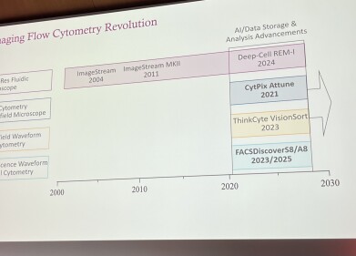
To address this, the group evaluated a prototype three-dimensional imaging flow cytometry platform capable of capturing whole-cell volumetric information. In proof-of-concept experiments, cells received a fluorescently labelled oligonucleotide and were monitored over time. Segmentation defined lysosome-like compartments, and signal outside this mask was quantified as a ratio. Newman highlighted practical constraints, including lysosomal tracker dependence on acidity and potential fixation artefacts, and stressed the need for careful validation. Nonetheless, early data suggested measurable temporal shifts, including small reductions in lysosome-associated signal at later time points.
She broadened the discussion to mass spectrometry-based methods and the value of combining imaging with chemical depth. Secondary ion mass spectrometry – long used in materials science – is increasingly applicable to questions in biology. Each pixel yields a mass spectrum, enabling extraction of specific chemical fragments and depth profiling through cellular layers. Across datasets, compound uptake varied markedly between cells, reinforcing the dominance of heterogeneity.
This variability motivated a move into single-cell proteomics. Workflows combine cell isolation or sorting with high-sensitivity mass spectrometry, accessed internally or through collaboration with national measurement laboratories. In a case study using amiodarone, induced pluripotent stem cell-derived macrophages showed dose-dependent lysosomal enlargement and pronounced heterogeneity. Sorting based on lysosomal fluorescence followed by proteomics recovered thousands of proteins per condition and revealed profiles consistent with phospholipidosis biology.
She concluded that cellular heterogeneity is signal, not noise. Microscopy shows where events occur, while mass spectrometry explains how they arise. Together, these approaches enable stronger assays, more predictive models and therapies that achieve meaningful target access at realistic doses.


Digital Edition
Lab Asia Dec 2025
December 2025
Chromatography Articles- Cutting-edge sample preparation tools help laboratories to stay ahead of the curveMass Spectrometry & Spectroscopy Articles- Unlocking the complexity of metabolomics: Pushi...
View all digital editions


Events
Jan 21 2026 Tokyo, Japan
Jan 28 2026 Tokyo, Japan
Jan 29 2026 New Delhi, India
Feb 07 2026 Boston, MA, USA
Asia Pharma Expo/Asia Lab Expo
Feb 12 2026 Dhaka, Bangladesh


