-
 Phantom midge (Chaoborus) larva,
 -
 A view across a CCD
 -
 Supramolecular Nanoring Network,
 -
 Titanium 'Boulder Field',
News
RMS Scientific Imaging Competition 2015 - Winners Announced
Aug 18 2015
From tiny mites to titanium ‘boulders’ the Royal Microscopical Society have announced images that are both stunning and technically challenging, that have taken the top spots in this year’s internationally-acclaimed Scientific Imaging Competition.
The winners were announced with prizes presented at the RMS’ flagship event, the Microscience Microscopy Congress in Manchester, where visitors were also able to view the display of all the Competition’s shortlisted images.
Electron Microscopy, Life Sciences
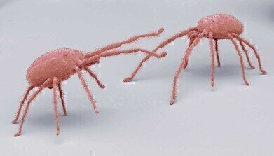
1st – Steve Gschmeissner - Mighty mites SEM Image, Coloured scanning electron micrograph (SEM) of predatory mites. Mites are small arthropods belonging to the subclass Acari and the class Arachnida. Mites are among the most diverse and successful of all the invertebrate groups.
2nd – Yuan-Chih Chang and Silk Yu Lin, Academia Sinica –The marine bacterium, Simiduia agarivorans
Electron Microscopy, Physical Sciences
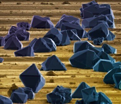
1st – Paul Gunning, Smith & Nephew Research Centre - Titanium 'Boulder Field', Electrochemically produced micro-crystals of titanium deposited on a machined titanium alloy surface give the appearance of a boulder field.
2nd – Kunwu Fu, Nanyang Technological University - Micro-tree of MAPbI3 perovskite, The image shows the film microstructures of methylammonium lead iodide perovskite material after being poled by high electric field.
Light Microscopy, Life Sciences
1st – David Linstead - Phantom midge (Chaoborus) larva, Live phantom midge larva imaged with a Zeiss X4 planapo objective on a Zeiss Standard trinocular microscope.
2nd – Eva Wegel, John Innes Centre – Chloroplast, This image shows chlorophyll autofluorescence in a live (dividing) chloroplast isolated from Arabidopsis thaliana leaves.
Light Microscopy, Physical Sciences
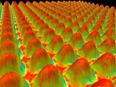
1st – Kevin Smith, MetPrep Ltd– A view across a CCD, An image of a camera chip when viewed at 1000x using an Olympus LEXT OLS4000 Laser Scanning Confocal Microscope
2nd – Claire Trease, Kingston University - A thin polymer film patterned with micro-pillars (dark circles) formed by an electrohydrodynamic instability and viscous fingers (large circle) formed from a Saffman-Taylor instability, transmitted light 500x.
Scanning Probe Microscopy
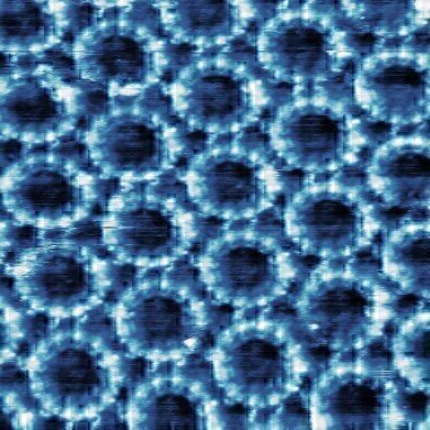
1st – Alex Summerfield, University of Nottingham - Supramolecular Nanoring Network, Constant current Scanning Tunnelling Microscopy image of a porphyrin nanoring network on a HOPG surface imaged in liquid.
2nd – Dipesh Khanal, University of Sydney – Human Tooth Enamel, Lorentz Contact Resonance microscopy image of human tooth enamel; prism (Red color,higher stiffness) and surrounding organic sheath (yellow color,lower stiffness)
Short Video
The winners of the short video category were:
1st – Michael Weber, Max Planck Institute of Molecular Cell Biology and Genetics - The Zebrafish Cardiovascular System, Recording of a four day old zebrafish embryo expressing fluorescent proteins for vasculature (upper panel / cyan), heart muscle and blood cells (lower panel / red).
2nd – Gopi Shah, Max Planck Institute of Molecular Cell Biology and Genetics– Zebrafish Development, A developing zebrafish embryo from 5 to 19 hours post fertilisation is shown in the movie. Every cell in the embryo is labelled with a nuclear marker and is colour coded for its position along the depth of the sample.
The next RMS Scientific Imaging Competition will take place in 2017, submissions are already being invited at www.rms.org.uk/imagingcomp2017


Digital Edition
Lab Asia Dec 2025
December 2025
Chromatography Articles- Cutting-edge sample preparation tools help laboratories to stay ahead of the curveMass Spectrometry & Spectroscopy Articles- Unlocking the complexity of metabolomics: Pushi...
View all digital editions


Events
Jan 21 2026 Tokyo, Japan
Jan 28 2026 Tokyo, Japan
Jan 29 2026 New Delhi, India
Feb 07 2026 Boston, MA, USA
Asia Pharma Expo/Asia Lab Expo
Feb 12 2026 Dhaka, Bangladesh


