-
 Macro to micro fluorescent in vivo imaging with the iBox Explorer Imaging Microscope
 -
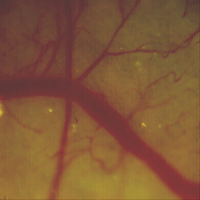
 Magnification at 8.8x of a skin-flap. Migrated dual-colored HT-1080 cells (bright yellow) within a distal vessel have begun to extravasate. The field of view corresponds to a 1.7 x 1.7mm window.
 -
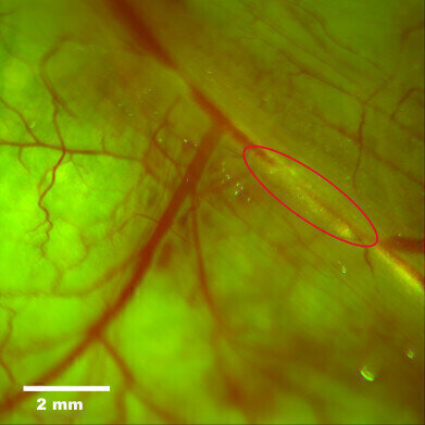
 A multiplexed image of a skin flap, 2.5x. The major vessel in the field corresponds to the epigastrica cranialis vein of a nude mouse. Clusters of dual-colored HT-1080 cells can be visualized moving through the vasculature in the right-most aspect of the field. The field of view is 5.8 x 5.8mm.
 -
 UVP - Upland, California and Cambridge UK
Microscopy & microtechniques
Macro to Micro Fluorescence In Vivo Imaging with the iBox Explorer Imaging Microscope
Jun 07 2012
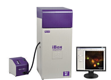
The iBox® Explorer™ Imaging Microscope combines enables brilliant resolution macro to micro technology for in vivo animal fluorescent imaging. The iBox Explorer provides breakthrough advances in its dual lighting system and software controlled objectives for imaging of tissues, tissue margins and individual cells.
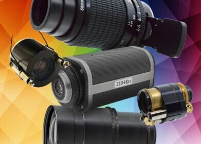
"The iBox Explorer is significant for speed and versatility," according to Sean Gallagher, VP and CTO of UVP. "Enabling the rapid and multiplexed fluorescence detection of tumor margins and micro metastasis, the Explorer cleanly separates normal from cancer tissues via the cell's fluorescent signature. Operating in the visible and NIR wavelengths, the Explorer yields detailed images of tissues and cells or, using the joy stick, 'flies' across an area such as the open abdominal region or skin flap of a mouse for rapid screening. In addition to imaging both the whole organ and cells of small animals, the Explorer delivers optical configurations that are parcentered and parfocal, allowing seamless imaging through the magnification ranges. The leading-edge cooled color camera enables quick detection, image capture and high throughput. The software automates research with total system control and allows easy creation of templates for reproducible and consistent results."
The iBox Explorer Fluorescence Microscope is the ideal system for applications including:
- Tumor shedding and angiogenesis
- Micro/Macro metastases
- Tumor/host margins and interactions
- Tumor micro environment
- Primary tumor growth
- Hematogenous and Intralymphatic trafficking
- Extravasation
A key component of the iBox Explorer is the BioLite™ Xe light source which provides a bright illumination source for multispectral fluorescent, visible, and NIR excitation. The BioLite Xe source houses a xenon lamp that allows brilliant excitation of fluorescent probes. The BioLite Xe includes a motorized filter wheel for the addition of up to eight independent excitation filters and allows for convenient switching between experiments and multiplexing applications. GFP/RFP filter sets are included and additional filters are available. View a selection of excitation and emission filters available or talk to a UVP BioImaging Specialist for further filter or system details.
Alex Waluszko, VP for Marketing/Sales, reports "Our research identifies the requirement for quick viewing of fluorescent markers from whole organ to single cell was an unmet need. The iBox Explorer supplies an economical solution for even the smallest labs and budgets in the cancer research field."
For more information on the iBox Explorer applications, see UVP's Application Notes.
Digital Edition
Lab Asia Dec 2025
December 2025
Chromatography Articles- Cutting-edge sample preparation tools help laboratories to stay ahead of the curveMass Spectrometry & Spectroscopy Articles- Unlocking the complexity of metabolomics: Pushi...
View all digital editions
Events
Jan 21 2026 Tokyo, Japan
Jan 28 2026 Tokyo, Japan
Jan 29 2026 New Delhi, India
Feb 07 2026 Boston, MA, USA
Asia Pharma Expo/Asia Lab Expo
Feb 12 2026 Dhaka, Bangladesh
.jpg)
-(2).jpg)


