-
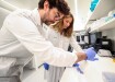
 Video microscopy was used to obtain results
Microscopy & microtechniques
Microscopy used in cell division project
Apr 06 2010
A six-year study that used microscopy to examine genetic cell division has now been published.
Findings from the MitoCheck project, coordinated by the Research Institute of Molecular Pathology (IMP) in Vienna, were outlined in the Science and Nature journals.
According to IMP, a facility founded in 1988, the process of cell division has "puzzled biologists for the past 150 years" and the work of researchers led by Jan-Michael Peters has come closer to solving the riddle.
Experts had to inactivate more than 22,000 genes in cultured cells in order to discover which ones were important for this process.
Video microscopy was then utilised to make movies of the samples to see whether powering down the individual organism units affected division.
"Our database is going to be an important source of information for many areas of biomedical research," Dr Peters suggested, adding that cooperation between international departments enabled the venture to be a success.
.jpg)
-(2).jpg)

Digital Edition
Lab Asia Dec 2025
December 2025
Chromatography Articles- Cutting-edge sample preparation tools help laboratories to stay ahead of the curveMass Spectrometry & Spectroscopy Articles- Unlocking the complexity of metabolomics: Pushi...
View all digital editions


Events
Jan 21 2026 Tokyo, Japan
Jan 28 2026 Tokyo, Japan
Jan 29 2026 New Delhi, India
Feb 07 2026 Boston, MA, USA
Asia Pharma Expo/Asia Lab Expo
Feb 12 2026 Dhaka, Bangladesh


