-
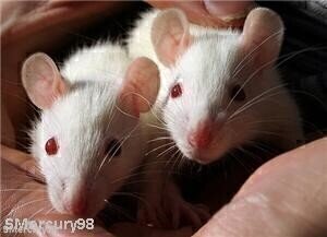 The skin was grafted onto mice
Microscopy & microtechniques
Microscopy methods developed in fake skin experiment
Apr 22 2010
Laboratory scientists have developed new microscopy methods in order to examine fake tissue surgically added to mice.
Researchers at the University of Granada needed to introduce several inmunofluorescence techniques to enable experiments on the skin, Diligent Media Corporation reports.
The substitute membranes, created using tissular engineering, underwent quality control operations - including checks for cell proliferation, the expression of cytokeratin and the presence of differentiating morphological markers.
Following tests, the lab experts found satisfactory biocompatibility levels with the mice, with the skin showing no signs of infection or rejection.
According to the news provider, all of the animals used in the study exhibited granulation of the grafted tissue within six days, while nearly three weeks later cicatrisation - the forming of scars - had completed.
Earlier this month, the Engineer magazine reported that the latest microscopy procedures are being implemented in a Wellcome Laboratories study to develop more effective treatments for eye diseases.
.jpg)
-(2).jpg)


Digital Edition
Lab Asia Dec 2025
December 2025
Chromatography Articles- Cutting-edge sample preparation tools help laboratories to stay ahead of the curveMass Spectrometry & Spectroscopy Articles- Unlocking the complexity of metabolomics: Pushi...
View all digital editions


Events
Jan 21 2026 Tokyo, Japan
Jan 28 2026 Tokyo, Japan
Jan 29 2026 New Delhi, India
Feb 07 2026 Boston, MA, USA
Asia Pharma Expo/Asia Lab Expo
Feb 12 2026 Dhaka, Bangladesh


