-
-
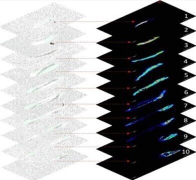 Ten consecutive tissue sections from a single C. elegans nematode, shown as optical images (left stack) and corresponding lipid distribution maps generated by MALDI mass spectrometry imaging (MSI) (right stack). This sectional data enabled 3D reconstruction of lipid localization, highlighting organ-specific fat distribution along the body axis. Credit: Sara Mandic from Okayama University, Japan
Research news
Microfluidics and mass spectrometry maps the distribution of lipids in C. elegans with high precision
Sep 15 2025
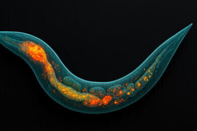
Understanding how fat molecules are organised and function in living organisms is essential to reveal mechanisms of ageing, disease, and metabolism. Caenorhabditis elegans –a transparent roundworm – has long served as a model to investigate fat storage because of its genetic similarity to humans and its well-defined anatomy. Yet the visualisation of lipids at high resolution in such a small organism has remained a major technical obstacle.
A team at Okayama University in Japan, led by Professor Masazumi Fujiwara and doctoral researcher Ms Sara Mandic, working with Professor Ron M. A. Heeren of Maastricht University in the Netherlands, has reported a microfluidics-based workflow that enabled high-resolution, three-dimensional lipid imaging in C. elegans.
The researchers combined matrix-assisted laser desorption/ionisation mass spectrometry imaging (MALDI-MSI) with traditional lipid staining to identify and locate fat molecules inside the worm. To retain the internal anatomy, young adult nematodes were aligned and immobilised on a microfluidic chip, embedded in a gelatin–carboxymethyl cellulose mixture, cryosectioned, and analysed with MALDI-MSI. Each section was also stained with Oil Red O – a synthetic fat-soluble dye used to stain lipids in tissues – to highlight neutral fats, providing a complementary confirmation of the imaging results.
“This is the first time we have been able to map lipid distributions in C. elegans with such spatial resolution while preserving internal structures,” said Ms Mandic.
Conventional methods to study lipids have required compromises, either to stain fats without identifying them or to measure them without spatial context. The reported technique achieved both, by detecting individual lipid molecules and locating them within the anatomy.
“Our technique gave researchers a reliable way to study fat dynamics in specific tissues of a single nematode,” explained Ms Mandic.
The group identified lipids that clustered in distinct regions, such as the pharynx, intestine and reproductive system. One lipid associated with cholesterol metabolism was concentrated in the pharynx and anterior intestine, which suggested a possible role in nutrient absorption. Because the workflow preserved tissue structure, the results provided insight into how fats are organised and function across the worm’s body.
The study also moved beyond two-dimensional imaging. By aligning and stacking sequential slices, the team produced three-dimensional reconstructions of nematodes that revealed whole-body lipid distribution with striking anatomical detail. The reproducibility of the method was high, with variation between worms greater than any technical inconsistency.
“This method allowed us to see not just what lipids were present but exactly where they were inside the body. Whether [that was] in the intestine, pharynx, or embryos,” Ms Mandic added.
C. elegans shares many biological pathways with humans and the implications of the work are broad. The technique has provided researchers with the means to study lipid behaviour in response to genetic mutations, environmental stress, drug treatments and ageing – all critical to understanding human health. The group intends to apply the workflow to C. elegans strains that carry disease-related mutations and to combine it with quantitative lipid analysis.
“Our work opened the door to visualise lipid biology in an entirely novel way – one that is precise, reproducible, and rich in detail,” concluded Ms Mandic.
For further reading please visit: 10.1038/s41598-025-09577-9


Digital Edition
Lab Asia Dec 2025
December 2025
Chromatography Articles- Cutting-edge sample preparation tools help laboratories to stay ahead of the curveMass Spectrometry & Spectroscopy Articles- Unlocking the complexity of metabolomics: Pushi...
View all digital editions


Events
Jan 21 2026 Tokyo, Japan
Jan 28 2026 Tokyo, Japan
Jan 29 2026 New Delhi, India
Feb 07 2026 Boston, MA, USA
Asia Pharma Expo/Asia Lab Expo
Feb 12 2026 Dhaka, Bangladesh


