
Fluorescence
What is Super Resolution Fluorescence Microscopy?
Oct 24 2014
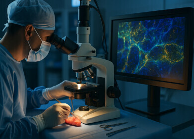
The ability to see an object, no matter how small, greatly enhances our capability to both describe and understand its appearance and behaviour. Biochemists need to view cells, viruses and protein molecules to allow them to describe and predict how our bodies might behave. To do this they have to use radiation that doesn’t harm or destroy the objects they are investigating, and this means using radiation in the visible range and using optical microscopy.
Optical Microscopy — a limited success
Scientists have been using optical microscopy since the seventeenth century to study the human body. Improvements in optics over the centuries have enabled better and smaller images to be seen, but the resolution of standard optical microscopes is limited by the very feature that makes it important to biochemists — light.
Although it is possible to arrange lenses and optical elements so that an image can be made as big as we like, this doesn’t mean that the finer and finer details can still be seen. Visible light, and other parts of the electromagnetic spectrum, behave as particles and waves — the wave-particle duality of the quantum world. Waves can exhibit a property called diffraction, caused when waves are incident on an aperture or edge of a barrier, causing interference patterns in the waves. These interference patterns can combine to give a continuous interference pattern known as diffraction. And the amount of diffraction sets the limit on the resolution.
Limits of Vision
For many years the limit of detection of optical microscopes has been fixed by the wavelength of light at about 0.2 micrometres, and no amount of increased magnification could make this any smaller. With a limit of 0.2 µm, scientists can see down to cell level, and even see some of the larger cell structures such as the mitochondria. But interactions between smaller objects and the cell were impossible to see. This problem was overcome by several researchers in the last decades of the twentieth century — earning them the 2014 Nobel Prize in Chemistry.
Super Resolution Microscopy
The researchers developed optical microscopy techniques that allowed them to overcome the diffraction limit imposed when using light. Fluorescent microscopy was already used in biomedical research — with fluorescent molecules used to image parts of a cell. But the resolution was still too poor to see individual molecules or DNA strands. The Nobel Prize winners overcame this issue in different ways — independent of each other. From the use of lasers that caused parts of a molecule to fluoresce in-sequence — to turning a single molecule’s fluorescence on — the breakthroughs have allowed high resolution images of features never before seen in optical microscopes. This has meant that for the first time scientists can see right to the heart of a cell and its interactions. For details of how these techniques are being used read: Spinning Disk Super-Resolution Microscopy – Bringing Super-Resolution in Focus for the Cell Biologist.
-(3).jpg)

Digital Edition
Lab Asia Dec 2025
December 2025
Chromatography Articles- Cutting-edge sample preparation tools help laboratories to stay ahead of the curveMass Spectrometry & Spectroscopy Articles- Unlocking the complexity of metabolomics: Pushi...
View all digital editions


Events
Jan 21 2026 Tokyo, Japan
Jan 28 2026 Tokyo, Japan
Jan 29 2026 New Delhi, India
Feb 07 2026 Boston, MA, USA
Asia Pharma Expo/Asia Lab Expo
Feb 12 2026 Dhaka, Bangladesh


