-
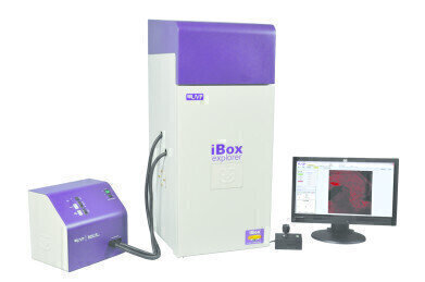 UVP's iBox® Explorer (2) Imaging Microscope.
 -
-

Microscopy & microtechniques
Imaging of Fluorescent Metastatic Mouse Models of Pancreatic Cancer - Using the iBox Explorer2 Imaging Microscope
Oct 02 2013
Cancer metastases are responsible for the majority of cancer-related deaths. Approximately 50% of cancer patients die within five years as a result of cancer-related problems, mostly attributable to metastatic lesions caused by the spread of cancers to near and distant sites. A hypothesis is that cancer metastasis arises from rare cells in the primary tumor that acquire the ability to progress through sequential steps necessary to grow at a distant site. Various mouse model systems are used to experimentally recapitulate and dissect the various steps of the metastatic process.
To visualise and track metastatic process, an imaging system with sensitive signal detection is important to the study. Here, UVP's iBox® Explorer2 Imaging Microscope reveals patterns of metastasis formation by human pancreatic carcinoma cells in mice. iBox Explorer2 quickly detects low fluorescent signals with its high resolution, high sensitivity camera. Excitation/emission filter combinations detect various fluorescent proteins and maximise emission intensity while reducing unwanted photons from auto fluorescence or bleed-through by other fluorophores.
Human pancreatic cancer cell line MIA-PaCa-2 was transduced with RFP-tagged plasmid. Cell culture and selection of MIA-PaCa-2 cells stably expressing RFP were conducted. A six-week-old male nude mice (BALB/c-nu/nu) was acclimated for one week and MIA-PaCa-2 cells stably expressing RFP were harvested. Cells (1 × 10⁷ cells in 0.1 mL PBS) were subcutaneously injected into the nude mice.
Ten weeks after MIA-PaCa-2-RFP inoculation, mice were sacrificed and organs dissected out. Images of mice organs, including spleen, kidney, intestine, heart and liver, were captured with the iBox Explorer - configured with RFP excitation (525BP45) and emission (650BP50) filters. The system incorporates a Xenon light source for sample illumination, capable of emitting high intensity excitation in the visible to NIR wavelengths.
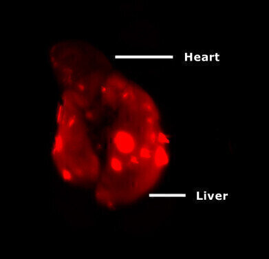
The dissected organs from the mouse inoculated with MIA-PaCa-2-RFP cells and control mouse were imaged using the iBox Explorer to detect the RFP signal. Liver and heart from the inoculated mouse were dissected. At 0.5x mangification, strong RFP signal was detected from liver, but not from heart or any other tissues (Fig. 2). Ex vivo imaging of the excised organs from MIA-PaCa-2-RFP inoculated mouse. Strong RFP signal was detected from the liver, but not from the heart. There was no RFP signal observed in any organ tissues in the control mouse.
Preliminary results showed metastasis formation in the liver 10 weeks after the human pancreatic cancer cell was inoculated in the mouse. This application illustrates iBox Explorer's functionality in imaging fluorescently-labeled tumor tissue. Using the system's high-sensitivity, cooled CCD camera and wide spectra filters, images with clear fluorescent signals and tissue structure were obtained. iBox Explorer evaluates and monitors metastatic process in various preclinical animal models. It can make in vivo and ex vivo imaging in mouse models easier, faster, and more accurate. The system's large selection of excitation/emission filters enables researchers to visualise multiple fluorescent signals in the same model.
Watch the webinar to learn more about dynamic in vivo imaging applications.
.jpg)
-(2).jpg)

Digital Edition
Lab Asia Dec 2025
December 2025
Chromatography Articles- Cutting-edge sample preparation tools help laboratories to stay ahead of the curveMass Spectrometry & Spectroscopy Articles- Unlocking the complexity of metabolomics: Pushi...
View all digital editions


Events
Jan 21 2026 Tokyo, Japan
Jan 28 2026 Tokyo, Japan
Jan 29 2026 New Delhi, India
Feb 07 2026 Boston, MA, USA
Asia Pharma Expo/Asia Lab Expo
Feb 12 2026 Dhaka, Bangladesh


