Microscopy & microtechniques
Innovative technology opens up new biomolecule analysis possibilities
Feb 26 2014
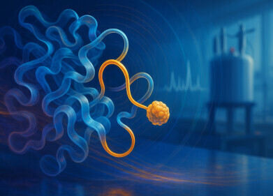
At the German Electron Synchnotron Facility (DESY) in Hamburg, scientists from the Centre for Free-Electron Laser Science (CFEL), the European Molecular Biology Laboratory (EMBL) and the universities of Hamburg and Lübeck have created new analysis possibilities for delicate biomolecules. At the X-ray source PETRA III, the scientists X-rayed micrometre-sized crystals of a key enzyme involved in sleeping sickness, thereby determining its atomic structure. They obtained structural data correspond to earlier analyses of the same enzyme at the world’s most powerful X-ray laser in Stanford (US). “Since only a few of these valuable crystals are needed, this method opens up new possibilities for the structural analysis of biomolecules,” said CFEL scientist Cornelius Gati, lead author of a research study* which describes this new method.
Through the use of x-ray crystallography biologists can determine a molecules functioning and properties by decoding its structure. In contrast to other materials however, many biomolecules are difficult to crystallise. “It requires a considerable effort to grow large enough crystals from complicated proteins for standard analyses,” said Lars Redecke from the joint Laboratory for Structural Biology of Infection and Inflammation of the universities of Hamburg and Lübeck. Some biomolecules at best form microcrystals which until now could only be analysed with the extremely intense light of X-ray lasers at large scale facilities.
At DESY’s X-ray source PETRA III, the team has now found a way to investigate microcrystals with more common research light sources as well – with the so-called synchrotron radiation sources. They grew microcrystals typically 4 to 15 thousandths of a millimetre from the enzyme cathepsin B of the Trypanosoma bruceiparasite, the cause of sleeping sickness, which were then scanned using the fine and intense X-ray beam of DESY’s synchrotron radiation source PETRA III operated by the European Molecular Biology Laboratory (EMBL).
With this method, the scientists were able to determine from 80 crystals the atomic structure of the enzyme with a spatial resolution of 0.3 millionth of a millimetre (nanometre). With a volume of 10 cubic micrometres, these crystals are among the smallest protein crystals that have been analysed with synchrotron radiation,” EMBL scientist Thomas Schneider points out.
“Our method is especially interesting for difficult to crystallise molecules,” said Redecke. “An advantage of this procedure is that we can work with components that are existing or will be available in the future at many research light sources,” explains Gleb Bourenkov from EMBL, the second lead author of the study.
“This approach will complement X-ray laser studies of difficult biological samples, allowing researchers to make optimal use of facilities that are in high demand,” said CFEL scientist Henry Chapman. Since this technology is also valuable for other research light sources, several research centres have already expressed their interest.
Reference: "Serial crystallography on in vivo grown microcrystals using synchrotron radiation"; Cornelius Gati, Gleb Bourenkov, Marco Klinge, Dirk Rehders, Francesco Stellato, Dominik Oberthür, Oleksandr Yefanov, Benjamin P. Sommer, Stefan Mogk, Michael Duszenko, Christian Betzel, Thomas R. Schneider, Henry N.Chapmana, and Lars Redecke; IUCrJ (February 2014)
Digital Edition
Lab Asia Dec 2025
December 2025
Chromatography Articles- Cutting-edge sample preparation tools help laboratories to stay ahead of the curveMass Spectrometry & Spectroscopy Articles- Unlocking the complexity of metabolomics: Pushi...
View all digital editions
Events
Jan 21 2026 Tokyo, Japan
Jan 28 2026 Tokyo, Japan
Jan 29 2026 New Delhi, India
Feb 07 2026 Boston, MA, USA
Asia Pharma Expo/Asia Lab Expo
Feb 12 2026 Dhaka, Bangladesh

.jpg)
-(2).jpg)


