
Research news
Scientists observe protein clusters in human brain tissue for first time that are thought to trigger Parkinson’s disease
Oct 21 2025
For the first time researchers from UK’s Cambridge University, University College London and Francis Crick Institute, alongside Canada’s Polytechnique Montréal have directly visualised alpha-synuclein oligomers in human brain tissue. The breakthrough offers vital insight into how Parkinson’s disease begins and spreads
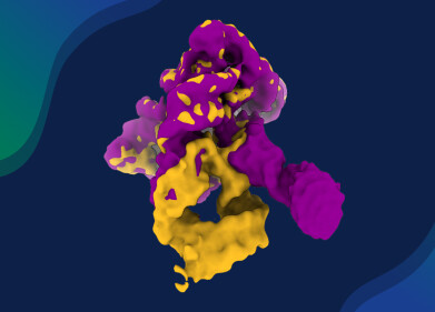
Scientists have – for the first time – directly visualised and quantified the protein clusters thought to initiate Parkinson’s disease in human brain tissue, marking a major advance in understanding this fast-growing neurological disorder. The science community has long considered the small protein clusters known as alpha-synuclein oligomers most likely to be the culprits behind the onset of Parkinson’s disease.
However, these structures have long evaded direct detection in human brain tissue, until the team constituted from the University of Cambridge, University College London, the Francis Crick Institute and Polytechnique Montréal, Canada, developed a method to observe them. The new imaging technique enables scientists to see, count and compare oligomers in post-mortem human brain samples. One of the researchers described this capability as ‘like being able to see stars in broad daylight.’
The study offers fresh insight into how Parkinson’s spreads through the brain and could support the development of tools which would be useful in earlier diagnosis and also potential treatments. Approximately 166,000 people in the UK live with Parkinson’s, with this number is rising. And worldwide, the number of people affected is expected to double to 25 million by 2050. Although existing medication can relieve symptoms such as tremor and stiffness, there is currently no treatment which slows or stops the disease.
For more than a century, doctors have identified Parkinson’s disease by the presence of large protein deposits known as Lewy bodies. However, scientists have long suspected that smaller, earlier-forming oligomers may be responsible for the initial damage to brain cells. Until now, these oligomers have been too small to see, measuring only a few nanometres in length.
“Lewy bodies are the hallmark of Parkinson’s but they essentially tell you where the disease has been, not where it is right now,” said Professor Steven Lee from Cambridge’s Yusuf Hamied Department of Chemistry, who co-led the study.
“If we can observe Parkinson’s at its earliest stages, that would tell us a whole lot more about how the disease develops in the brain and how we might be able to treat it,” he added.
The team’s technique, called Advanced Sensing of Aggregates for Parkinson’s Disease (ASA-PD), uses ultra-sensitive fluorescence microscopy to detect and analyse millions of oligomers in post-mortem brain tissue. Because the oligomers are extremely small, their optical signal is faint. ASA-PD enhances that signal while reducing background interference, dramatically increasing sensitivity until individual alpha-synuclein oligomers can be observed and characterised.
“This is the first time we have been able to look at oligomers directly in human brain tissue at this scale – it’s like being able to see stars in broad daylight,” said Dr Rebecca Andrews, who co-authored the study while a postdoctoral researcher in Lee’s laboratory.
“It opens doors in Parkinson’s research,” she added.
The researchers compared brain tissue from individuals who had Parkinson’s disease with tissue from healthy individuals of similar age. They found that oligomers exist in both groups, but those in the Parkinson’s samples were larger, brighter and more numerous, indicating a link to disease progression. They also identified a subclass of oligomers found only in the Parkinson’s brains, which could represent the earliest visible markers of the disease, potentially appearing years before symptoms develop.
“This method does not just give us a snapshot,” said Professor Lucien Weiss from Polytechnique Montréal, who co-led the research.
“It offers a whole atlas of protein changes across the brain, and similar technologies could be applied to other neurodegenerative diseases such as Alzheimer’s and Huntington’s.
“Oligomers have been the needle in the haystack, but now that we know where those needles are, it could help us target specific cell types in certain regions of the brain,” Weiss said.
The research was supported by Aligning Science Across Parkinson’s (ASAP), the Michael J. Fox Foundation and the Medical Research Council, part of UK Research and Innovation. The scientists expressed their gratitude to the patients, families and carers who donated tissue to brain banks, making the study possible.
For further reading please visit: 10.1038/s41551-025-01496-4


Digital Edition
Lab Asia Dec 2025
December 2025
Chromatography Articles- Cutting-edge sample preparation tools help laboratories to stay ahead of the curveMass Spectrometry & Spectroscopy Articles- Unlocking the complexity of metabolomics: Pushi...
View all digital editions


Events
Jan 21 2026 Tokyo, Japan
Jan 28 2026 Tokyo, Japan
Jan 29 2026 New Delhi, India
Feb 07 2026 Boston, MA, USA
Asia Pharma Expo/Asia Lab Expo
Feb 12 2026 Dhaka, Bangladesh


