Research news
AI tool gives doctors a head start in detecting skin cancer
Jun 19 2025
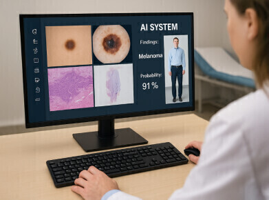
Detection of melanoma and other skin diseases could become faster and more accurate following the development of an artificial intelligence (AI) tool that simultaneously analyses multiple types of medical images. The system – developed by an international team led by Monash University in Melbourne – is one of the first AI models tailored for real-world dermatological practice.
The model, called PanDerm, integrates a range of visual inputs used in skin examinations, including close-up photographs, dermoscopic images, pathology slides and full-body images. In clinical evaluations, the tool was shown to improve the diagnostic accuracy of doctors identifying skin cancer by 11 per cent. Among non-dermatologist healthcare professionals, diagnostic performance across a wider array of skin conditions improved by 16.5 per cent.
PanDerm also demonstrated an ability to identify potentially cancerous lesions before they were detected by clinicians. Trained on over two million skin images collected from 11 institutions across multiple countries, the model incorporates four distinct imaging types commonly used in dermatology.
Associate Professor Zongyuan Ge, lead co-author of the study and an expert in AI and computer vision at Monash University’s Faculty of Information Technology, said current AI tools used in dermatology were often restricted to specific tasks, such as assessing dermoscopic images for skin cancer.
“Previous AI models have struggled to integrate and process various data types and imaging methods, reducing their usefulness to doctors in differing real-world settings,” said Associate Professor Ge.
“PanDerm is a tool designed to work alongside clinicians, helping them interpret complex imaging data and make informed decisions with more confidence,” he said.
Unlike earlier systems built for single diagnostic tasks, PanDerm has been evaluated across a broad spectrum of clinical applications. These include skin cancer screening, recurrence prediction, mole counting, lesion tracking, skin type assessment and the diagnosis of various skin diseases. The model also performed segmentation tasks to outline lesions within images.
Despite its complexity, PanDerm consistently delivered best-in-class results, even when trained on only five to 10 per cent of the annotated data typically required.
In clinical use, PanDerm functions as a diagnostic support tool. It processes multiple imaging modalities and produces probability-based diagnostic assessments to assist clinicians in interpreting findings. This multimodal capability is especially beneficial in primary care settings and in rural or regional locations, where access to dermatology specialists may be limited.
Siyuan Yan, a doctoral candidate in the Monash University Faculty of Engineering and first author of the paper, said that the system’s success lay in its ability to combine information from diverse sources.
“By training PanDerm on data from multiple imaging techniques, we have developed a system that mirrors how dermatologists think – by synthesising information from a variety of visual sources. This allows for a more holistic analysis of skin conditions than earlier AI tools limited to a single image type,” said Mr Yan.
Given that skin conditions affect approximately 70 per cent of the global population, the ability to identify them early is critical for improving treatment outcomes. Professor Victoria Mar, Director of the Victorian Melanoma Service at Alfred Health, in Victoria, Australia and a lead co-author, said PanDerm shows potential for enhancing the monitoring of lesions over time and revealing biological clues about disease progression.
“This kind of assistance could support earlier diagnosis and more consistent monitoring for patients at risk of melanoma. In hospitals and clinics, clinicians use diverse imaging methods to diagnose skin conditions. PanDerm is built to complement that approach,” said Professor Mar
Although the tool has shown promising results in research settings, PanDerm remains under evaluation prior to broader implementation in healthcare systems. The research team plans to develop more comprehensive assessment frameworks to evaluate its performance across a wider range of skin conditions and clinical scenarios.
The next phase will focus on standardising protocols for cross-demographic analysis and ensuring the model performs equitably across diverse populations and healthcare environments.
The study was led by AI and machine learning experts from the AIM for Health Lab at Monash University’s Faculty of Information Technology, in partnership with researchers and clinicians from Alfred Health, The University of Queensland, Princess Alexandra Hospital in Brisbane, Royal Prince Alfred Hospital, Melbourne, NSW Health Pathology, the Medical University of Vienna, NVIDIA AI Technology Centre in Singapore, the University of Florence and Hospital General Universitario de Alicante in Spain.
For further reading please visit: 10.1038/s41591-025-03747-y

Digital Edition
Lab Asia Dec 2025
December 2025
Chromatography Articles- Cutting-edge sample preparation tools help laboratories to stay ahead of the curveMass Spectrometry & Spectroscopy Articles- Unlocking the complexity of metabolomics: Pushi...
View all digital editions


Events
Jan 21 2026 Tokyo, Japan
Jan 28 2026 Tokyo, Japan
Jan 29 2026 New Delhi, India
Feb 07 2026 Boston, MA, USA
Asia Pharma Expo/Asia Lab Expo
Feb 12 2026 Dhaka, Bangladesh


