Laboratory products
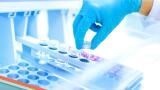
Protein gel electrophoresis followed by common staining protocols is a widely accepted technique for analysing specific proteins in complex samples. Generally, proteins separated by electrophoresis are visualised using staining and time-consuming de-staining procedures.
Imaging of proteins without staining is a new technique which is coming into the spotlight as a result of numerous advantages over traditional staining practices. This methodology is based on gel chemistry where the gels contain a trihalocompound which covalently binds to the amino acid tryptophan in protein samples when exposed to UV and subsequently produces fluorescence. This technique eliminates staining and time consuming de-staining steps to make experimental protocols extremely simple and time efficient.
Materials and Methods
MagicMark™ XP Western Protein Standard (Life Technologies) was separated on a 12.5% Fluorescent Sprint Next Gel (AMRESCO) for 15 minutes at 200V until the dye reached the bottom of the gel. The Next Gel running buffer was used for the separation of proteins in the gel. Fluorescent Sprint Next Gels are imaged and analysed in 15-20 minutes with the GelMax Imager, compared to 2-3 hours with traditional protein gel staining methods.
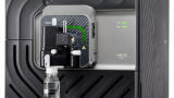
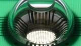
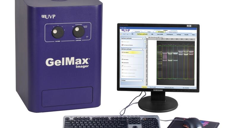
The GelMax Imaging System (Figure 1) was used to visualise the gel. The gel was exposed to two minutes of UV radiation from the system's built-in transilluminator. After UV exposure, an image of the gel was captured using a 10 second exposure time. An ethidium bromide emission filter was used for light optimisation. The image was then cropped and zoomed, and UVP’s VisionWorks®LS software used to study the unique characteristics of each individual band for quantitative analysis.
Results and Discussion
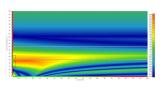
Figure 2 illustrates and validates the remarkable imaging capabilities of the GelMax Imaging System with Fluorescent Sprint Next Gels. This technique offers multiple advantages over traditional staining and normalization methods, including instant visualisation of protein bands after electrophoresis and UV exposure without the need for lengthy staining/de-staining steps. The sensitivity of this technique is comparable to typical staining methods such as Coomassie Blue staining.
Using the GelMax’s VisionWorksLS software, the relative amount of total protein in each lane on the gel can be measured and used for protein quantitation and normalisation.
Figure 2. Fluorescent Sprint Next Gel visualisation using the GelMax Imaging System
Conclusion
Fluorescent Sprint Next Gels can be conveniently imaged with the GelMax Imaging System by using the integrated UV light source and standard emission filters.
This technique remarkably improves standard protein detection protocols in terms of time efficiency and simplification of the procedure, allowing instant visualisation of gel separation and protein transfer.
For further information contact UVP.


.jpg)
-(2).jpg)