
Laboratory products
New Method for Non-Invasive Prostate Cancer Screening using a Confocal Raman Microscope Developed
Jan 06 2017
New research at the Guangdong Medical University suggests a laser-based approach could be the latest breakthrough in prostate cancer detection. The proposed non-invasive blood test uses a combination of two techniques: surface-enhanced Raman scattering (SERS) and a new mathematical analysis technique called support vector machine (SVM). Together, these techniques produce an accuracy up to 98.1%; a far cry from the relative guesswork of prostate-specific antigen (PSA) tests. Professor Shaoxin Li, the study leader at the University commented: “Compared to traditional screening methods, this method has the advantage of being non-invasive, highly sensitive, and very simple for prostate cancer screening.”
Professor Li continued: “Cancer is one of the diseases that seriously threatens human life. It is important to improve the survival of patients by early diagnosis and treatment. Currently, there are many diagnostic methods available—including B-mode ultrasound, CT scan, biopsy and histopathology assessment—but these techniques have various limitations. For example, B-mode ultrasound only discerns the solid tumour and is therefore not applicable to patients in the early stages of cancer. Biopsy and histopathology assessment are the gold standard of cancer examination but they are invasive and impractical for high-risk patients with multiple suspicious lesions. We hope to develop a rapid, non-destructive, optical diagnosis method to solve these problems.
“Advanced Raman spectroscopy has provided the possibility to meet our goals and after much evaluation of alternative Raman systems, including portable Raman instruments, we chose the Renishaw inVia confocal Raman microscope. We selected the inVia because it offers continuous scanning from 50 to 4000 wavenumbers [using SynchroScan, Renishaw’s patented method of acquiring wide-range spectra] and its high sensitivity makes it suitable for biological tissue measurement. It is also highly automated with software that is powerful and easy to use.”
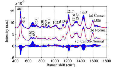
To illustrate the sensitivity in use, Professor Li showed an example comparing SERS spectra of serum samples with silver colloids. The differences in the spectra reveal the enormous potential to diagnose cancer using the serum SERS technique.
Results show normalised mean SERS spectra of prostate cancer and normal serum sample. (a) cancer, (b) normal, (c) difference in spectra (cancer-normal). shaded area represents the standard deviations.


Digital Edition
Lab Asia Dec 2025
December 2025
Chromatography Articles- Cutting-edge sample preparation tools help laboratories to stay ahead of the curveMass Spectrometry & Spectroscopy Articles- Unlocking the complexity of metabolomics: Pushi...
View all digital editions


Events
Jan 21 2026 Tokyo, Japan
Jan 28 2026 Tokyo, Japan
Jan 29 2026 New Delhi, India
Feb 07 2026 Boston, MA, USA
Asia Pharma Expo/Asia Lab Expo
Feb 12 2026 Dhaka, Bangladesh


