Fluorescence
In Vivo Fluorescence Imaging of Angiogenic Vessels in Liver Cancer Animal Model
Nov 06 2013
Angiogenesis is a vital component of both normal physiological processes and a number of disease states. Angiogenic process is a complex multistep phenomenon involving many growth factors and interactions between different cell types. In angiogenesis, there are two primary methods of vascular expansion where nutrient supply to tissues is adjusted to match physiological demand, one being the sprouting of new capillaries from existing blood vessels and another being vasculogenesis, the de novo generation of blood vessels. Vasculogenesis is an integral and essential component of embryonic development, and angiogenesis accompanies organ growth and regeneration.
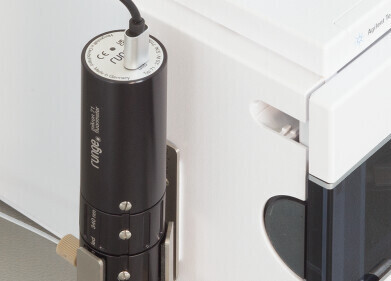
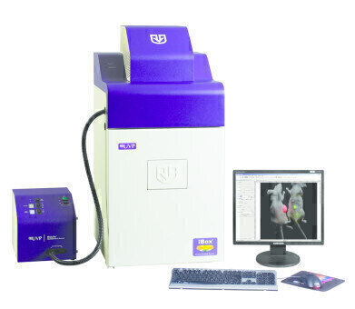
UVP’s iBox® Explorer2 Imaging Microscope provides an effective way to image angiogenesis in vivo, aiding researchers in understanding and evaluating different conditions that stimulate or inhibit angiogenesis in preclinical models and human disease. The iBox Explorer2 integrates a cooled, high resolution camera capable of detecting low fluorescent signals in angiogenesis across a wide spectral range within living animals. Here, the iBox Explorer2 is used to visualise angiogenesis vessels in the liver cancer animal model.
Materials and Methods
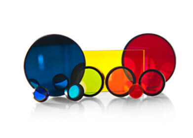
Hepatocellular Carcinoma Cell Line HepG2 was transfected with GFP-tagged plasmid. Six-week-old male nude mice were acclimated for one week. HepG2 cells stably expressing GFP were harvested and kept cold. Cells (1 × 10⁷ cells in 0.1 mL PBS) were subcutaneously injected into the mice. Three weeks after HepG2-GFP inoculation, the mice with tumor growths were selected and used for orthotropic transplantation. Tumor tissues were harvested and cut into pieces under aseptic conditions. Tumor pieces were orthotopically implanted into liver lobes. The mice were placed into cages and raised under Specific Pathogen Free (SPF) conditions. One month after tumor transplantation, mice were anesthetized and images captured with the iBox Explorer2 configured with GFP excitation/emission filters, 3.2MP camera and BioLite™ MultiSpectral Light Source.
Results
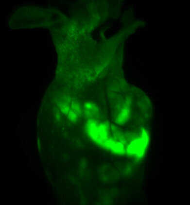
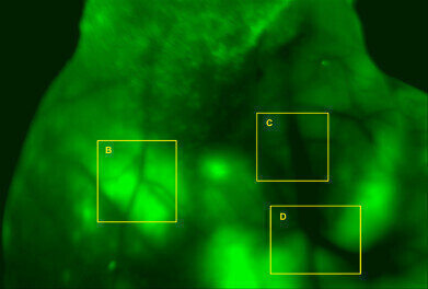
Images of GFP-fluorescence signals in nude mice were captured with the iBox Explorer2 at different magnification levels. GFP signals were detected in multiple areas of mice bodies, not only in the transplanted position, indicating that tumor cells had spread within the mice. In addition, images of angiogenic vessels around the tumor were captured.
Conclusion
In this experiment, strong fluorescence signals in mouse one month after tumor transplantation were captured. Tumor cell spread from liver lobes to other tissues inside the mouse body. To analyse angiogenic responses, angiogenic vessels around tumor tissues were visualised and images captured, numbers and lengths of vessels measured and recorded for further study. The experiment verified HepG2-GFP liver cancer mouse model is a reliable, reproducible system for angiogenesis study.
iBox Explorer2 Imaging Microscope is a valuable tool for preclinical in vivo angiogenesis imaging, making vessel imaging in mouse models easy and fast. The system enables high quality imaging techniques to monitor changes in vessels. This technique could be used for qualitative and quantitative assessment of various angiogenesis studies and monitoring antiangiogenic therapies, such as to understand regulating pathways or analyse angiogenic responses to specific perturbations of angiogenic process.
Read the full Application Note.
Digital Edition
Lab Asia Dec 2025
December 2025
Chromatography Articles- Cutting-edge sample preparation tools help laboratories to stay ahead of the curveMass Spectrometry & Spectroscopy Articles- Unlocking the complexity of metabolomics: Pushi...
View all digital editions
Events
Jan 21 2026 Tokyo, Japan
Jan 28 2026 Tokyo, Japan
Jan 29 2026 New Delhi, India
Feb 07 2026 Boston, MA, USA
Asia Pharma Expo/Asia Lab Expo
Feb 12 2026 Dhaka, Bangladesh
-(3).jpg)
