Chromatography
Scientists Rebuild Parts of the Human Heart
Sep 01 2019
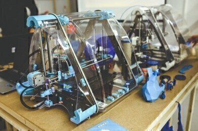
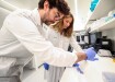
In a scientific breakthrough that could revolutionise cardiology, a team of researchers from Carnegie Mellon University in Pittsburgh has developed a 3D printer that uses collagen to manually reconstruct components of the human heart and build complex collagen scaffolds that replicate the look, feel and function of biological tissues.
As the main component of most tissues and organs in the human body, collagen is an incredibly versatile protein. Until now, it's been difficult to print living cells and soft biomaterials such as collagen because of the unstable nature. The new 3D printer developed by the Carnegie Mellon University overcomes this challenge with the use of a support gel. The gel allows the printer to create collagen-rich heart tissue and build parts of the organ, including capillaries, chambers, valves and vessels.
Introducing FRESH technology
The technique is known as FRESH, short for freeform reversible embedding of suspended hydrogels. A report published in the journal Science explains how the team overcame the challenge of 3D printing "soft and squishy" tissues by using a stabilising gel. "In our work we have solved this problem by 3D bioprinting inside a ‘support’ gel that prevents the cells and hydrogels (similar to gelatine) from collapsing,” explains study leader Adam Feinberg.
Armed with the new technique, Feinberg and his team were able to successfully 3D print collagen with the same ease as other materials such as metal and plastic. “Being able to do this means that we can now print scaffolds and tissues that actually have biological function,” he asserts.
Printing patient-specific anatomical structures
The method works by adding a supply of supportive gelatine microparticle slurry to the 3D printer. Bioinks containing unmodified collagen are then inserted into the printing needle, which creates thin, stacked layers of collagen and cells. After the printing process is complete, the room temperature is raised to 37°C which slowly melts the support gel and releases the printed collagen scaffold.
As well as general heart components, Feinberg also says FRESH can be used to print patient-specific anatomical structures using MRI or CT images. The images can then be used to generate unique 3D computer models and printing instructions that meet the exact needs of the patient.
If you like the concept of FRESH, you'll also find the revolutionary new LSER method fascinating. Find out more about how scientists are building an understanding of the interactions between analyte and sorbent by reading 'Characterisation of RP Sorbents by Linear Solvation Energy Relationships (LSER).'


Digital Edition
Lab Asia Dec 2025
December 2025
Chromatography Articles- Cutting-edge sample preparation tools help laboratories to stay ahead of the curveMass Spectrometry & Spectroscopy Articles- Unlocking the complexity of metabolomics: Pushi...
View all digital editions


Events
Jan 21 2026 Tokyo, Japan
Jan 28 2026 Tokyo, Japan
Jan 29 2026 New Delhi, India
Feb 07 2026 Boston, MA, USA
Asia Pharma Expo/Asia Lab Expo
Feb 12 2026 Dhaka, Bangladesh


