Optical imaging
Fluorescence Imaging Microscope Released
Oct 01 2010
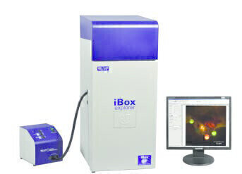
UVP, LLC exhibited at World Molecular Imaging Conference in Kyoto, Japan, to showcase the new iBox® Explorer™ Fluorescence Imaging Microscope along with UVP’s extensive line of iBox in vivo imaging systems for rapid high resolution analysis of fluorescently tagged cells, tissues, organs and whole animals in the visible and NIR wavelengths. UVP now offers three systems for real time, fluorescence imaging to speed up basic and preclinical research studies in tumours, oncology, heart and metabolic diseases.
The iBox Explorer is unique in its ability to micro image cells and organs subcutaneously and within the body cavity of living mice. The iBox Explorer joins the whole mouse imagers iBox Scienta and iBox Spectra in revolutionising in vivo fluorescent imaging. The iBox Explorer operates through its intuitive software control using optical configurations that are parcentered and parfocal, allowing seamless imaging through the magnification ranges. Included is a leading-edge high frame rate cooled color camera with technology that enables quick detection, image capture and high throughput. Applications of the iBox Explorer include imaging whole organs, tumor/host margins and interactions, tumour microenvironment, vasculature, and micro metastases.
UVP’s line of BioImaging Systems includes the BioSpectrum® multifunctional imaging systems which combines a high resolution CCD camera and advanced features for enhanced imaging fluorescent and chemiluminescent imaging capabilities.
.jpg)

Digital Edition
Lab Asia Dec 2025
December 2025
Chromatography Articles- Cutting-edge sample preparation tools help laboratories to stay ahead of the curveMass Spectrometry & Spectroscopy Articles- Unlocking the complexity of metabolomics: Pushi...
View all digital editions


Events
Jan 21 2026 Tokyo, Japan
Jan 28 2026 Tokyo, Japan
Jan 29 2026 New Delhi, India
Feb 07 2026 Boston, MA, USA
Asia Pharma Expo/Asia Lab Expo
Feb 12 2026 Dhaka, Bangladesh


