-
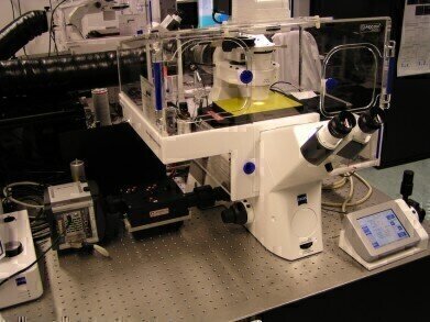 CLF's Octopus Imaging facility
News
Designer Nanomaterials Caught by Laser Octopus
Jun 29 2016
Researchers from the University of Bristol working with a team from the Science and Technology Facilities Council’s Central Laser Facility, UK have found a way of identifying two-dimensional nanomaterials, called platelet micelles, using the super resolution imaging of the STFC’s microscope facility ‘Octopus’.
Platelet micelles consisting of three concentric rectangles, each incorporating fluorescent dyes of a different colour and with a central hole, can be easily seen in a fluorescence microscope. However, because the rectangles are about 200 nm thick, they appear blurred and overlapping.
“A conventional microscope cannot resolve multicolour objects on this scale but the structured illumination microscope within ‘Octopus’ is ideally suited to imaging objects between 100 and 300 nanometres in size. These discoveries are the first use of super-resolution techniques in this type of materials science research. The work opens the doors to being able to image a whole range of new materials that previously could not be observed effectively at high resolution” said Dr Stephen Webb, from STFC’s Central Laser Facility (CLF).
Their research* reports that these micelles have a highly controllable structure and are easily assembled into larger structures.This, and the fact that they are easily functionalised, makes them a potential tool for a wider range of uses, including therapeutic applications and catalysis. For example, the circulation time of drug delivery vehicles in the body is dependent on their size and morphology. These features can be controlled in these micelles and the platelets can also be functionalised to contain medically relevant molecules.
Professor Ian Manners, who led the team from the University of Bristol, said “The characterisation using the super resolution imaging capability at the CLF was absolutely critical to the success of this work. Without the extra resolution that Octopus offered us, the internal structure of the micelles would not have been clear at all”.
The microscope used was funded by the Medical Research Council through a grant awarded to the Octopus group leader, Professor Marisa Martin-Fernandez, to develop super-resolution imaging for biomedical research. Ian Manners’ research is funded by both EPSRC and the European Research Council.
*Recently published in the journal Science


Digital Edition
Lab Asia Dec 2025
December 2025
Chromatography Articles- Cutting-edge sample preparation tools help laboratories to stay ahead of the curveMass Spectrometry & Spectroscopy Articles- Unlocking the complexity of metabolomics: Pushi...
View all digital editions


Events
Jan 21 2026 Tokyo, Japan
Jan 28 2026 Tokyo, Japan
Jan 29 2026 New Delhi, India
Feb 07 2026 Boston, MA, USA
Asia Pharma Expo/Asia Lab Expo
Feb 12 2026 Dhaka, Bangladesh


