-
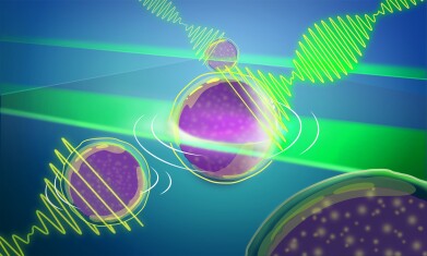 An artist's representation of the new Brillouin microscopy approach that allows entire light-sheets to interact with 3D biological samples. The scattered light reveals a unique optical interference signal that can be recorded with a custom-developed spectrometer, tremendously speeding up image acquisition. Credit: Credit: Daniela Velasco/EMBL
Microscopy & microtechniques
Pushing the limits of ‘custom-made’ Brillouin microscopy
Mar 21 2025
Advance to Brillouin microscopy by a European Molecular Biology Laboratory-engineered team widens the aperture and provides for quicker, more efficient 3D imaging of light-sensitive samples
The European Molecular Biology Laboratory (EMBL) has developed a novel methodology in microscopy which represents a 1,000-fold improvement in speed and throughput in Brillouin microscopy and provides a way to view light-sensitive organisms more efficiently.
“Over the years, we have progressed from being able to see just a pixel at a time to a line of 100 pixels, to now a full plane that offers a view of approximately 10,000 pixels,” said Dr. Carlo Bevilacqua, lead author the paper and an optical engineer in EMBL’s Prevedel team, in Heidelberg, Germany. “We were on a quest to speed up image acquisition.”
The technology is based on a phenomenon first predicted in 1922 by French physicist Léon Brillouin, who predicted that when light is shone on a material, it would interact with naturally occurring thermal vibrations within, exchanging energy and thereby slightly shifting the frequency – colour – of the light. Measuring this spectrum of the scattered light could therefore reveal information about the material’s characteristics.
Actual application of Brillouin Scattering microscopy only became possible much later, however, in the early 2000s. Technological advances now enabled measurement of tiny frequency shifts with high precision and sufficient throughput. This allowed them to compute mechanical properties of living biological samples. Initially scientists were only able to view one pixel at a time meaning that building up a worthwhile dataset was time-consuming, and hence severely limited the use of the microscopy method in biology.
In 2022, Bevilacqua and others in the EMBL Prevedel group were able to first expand the field of view to a line, and now with this latest development, to a full 2D field of view, which it turn also helps speed up 3D imaging.
“Just as the development of light-sheet microscopy marked a revolution in light microscopy because it allowed for faster, high-resolution, and minimally phototoxic imaging of biological samples, so too does this advance in the area of mechanical or Brillouin imaging,” said Dr Robert Prevedel, group leader and senior author on the paper.
“We hope this new technology – with minimal light intensity – opens one more ‘window’ for life scientists’ exploration,” he concluded.
For further reading please visit: 10.1038/s41566-025-01619-y
NOTE: EMBL – with 29 member states, the European Molecular Biology Laboratory has more than 110 independent research groups and service teams covering the spectrum of molecular biology across six sites in Barcelona, Grenoble, Hamburg, Heidelberg, EMBL-EBI Hinxton, and Rome.
.jpg)
-(2).jpg)

Digital Edition
Lab Asia Dec 2025
December 2025
Chromatography Articles- Cutting-edge sample preparation tools help laboratories to stay ahead of the curveMass Spectrometry & Spectroscopy Articles- Unlocking the complexity of metabolomics: Pushi...
View all digital editions


Events
Jan 21 2026 Tokyo, Japan
Jan 28 2026 Tokyo, Japan
Jan 29 2026 New Delhi, India
Feb 07 2026 Boston, MA, USA
Asia Pharma Expo/Asia Lab Expo
Feb 12 2026 Dhaka, Bangladesh


