-
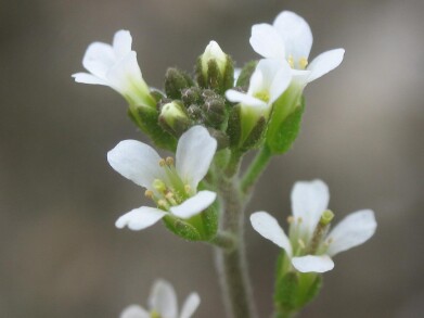 Arabidopsis was studied by the Rutgers' team. It is considered a 'model organism' with characteristics that are widely applicable in other organisms. Credit: Wikimedia Commons
Microscopy & microtechniques
Microscopy allows plants to be observed building cell walls for the first time
Mar 25 2025
Rutgers’ biophysicists, bioengineers and plant biologists capture first live images of cellulose construction leading to potential practical applications
A research team from Rutgers University-New Brunswick, New Jersey, USA, has captured images – over a continuous 24-hour period – of the synthesis of cellulose in living plant cell-walls. The work may lead to the development of more robust plants for the purpose of increased food production and lower-cost biofuels.
The dynamic process has not been observed before and adds to the fundamental knowledge of cell wall formation while increasing the range of practical applications from plant-derived products.
“This work is a direct visualisation of how cellulose synthesises and self-assembles into a dense fibril network on a plant cell surface. The study provides entirely new insights into how simple, basic physical mechanisms such as diffusion and self-organisation lead to the formation of complex cellulose networks in cells,” said Associate Professor Sang-Hyuk Lee, Department of Physics and Astronomy and an author of the study.
Three Rutgers’ laboratories, working across different academic disciplines, have collaborated for six years on the project. The team was drawn from the School of Arts and Sciences, the School of Engineering, and the School of Environmental and Biological Sciences.
Cellulose – chemically a carbohydrate – is the most abundant biopolymer on Earth and is the primary structural component of plant cell walls. It has many industrial applications for product manufacture including paper and clothing, but also has uses in filtration and as a food thickening agent.
The video images generated under a microscope show protoplasts from the Arabidopsis – a flowering plant from the brassica family. Protoplasts are cells with their walls removed. The images captured the process of the chaotic sprouting of cellulose fibre filaments that gradually self-assembled into a complex network on the cell’s outer surface. Arabidopsis has been extensively studied as a model organism.
“I was very surprised by the emergence of ordered structures out of the chaotic dance of molecules when I first saw these video images,” said Lee, who also is a faculty member at the Institute for Quantitative Biomedicine.
“I thought plant cellulose would be made in a lot more of an organised fashion, as depicted in classical biology textbooks.”
“This discovery opens the door for researchers to begin dissecting the genes that could play various roles for cellulose biosynthesis in the plant,” said Professor Eric Lam, Distinguished Professor in the Department of Plant Biology, another study author.
“[We will be able to] design better plants for carbon capture, improve tolerance to all kinds of environmental stresses, from drought to disease, and optimise second-generation cellulosic biofuels production.”
“I have always been fascinated by plants and how they capture sunlight via leaves into reduced carbon forms like cellulose that form cell walls. I [am] inspired by that experience to delve deeper into the fundamental phenomena of biomass production and [the] sustainable engineering [uses] to produce valuable bioproducts for societal benefit,” said study author Associate Professor Shishir Chundawat, Department of Chemical and Biochemical Engineering.
The team developed an advanced super-resolution – and minimally invasive technique – called total internal reflection fluorescence microscopy. The approach, which captured images only of the underside surface of cells, was sensitive enough to take videos for 24 hours without bleaching or destroying the cells.
Lee, a biophysicist with expertise in cutting-edge microscopy techniques for the study of living systems, oversaw the imaging efforts.
Chundawat led a team that pioneered a technique to allow scientists to tag the emerging cellulose tendrils with a fluorescent protein dye. Chundawat is a bioengineer and expert on protein engineering and glycoscience. To make the cells fluoresce to be detectable by the microscope, his team developed an engineered bacterial enzyme which binds specifically to cellulose.
Plant genetics and biotechnology expert, Lam, found a way to remove the cell wall of individual cells of Arabidopsis to create a ‘blank slate’ for new cell walls to be laid down by protoplast cells.
Other Rutgers scientists on the study included: Hyun Huh, a postdoctoral scientist with the Institute for Quantitative Biomedicine; Dharanidaran Jayachandran, a doctoral student, and Mohammad Irfan, a postdoctoral scientist in the Department of Chemical and Biochemical Engineering; and Junhong Sun, a lab technician in the Department of Plant Biology.
For further reading please visit: 10.1126/sciadv.ads6312
.jpg)
-(2).jpg)


Digital Edition
Lab Asia Dec 2025
December 2025
Chromatography Articles- Cutting-edge sample preparation tools help laboratories to stay ahead of the curveMass Spectrometry & Spectroscopy Articles- Unlocking the complexity of metabolomics: Pushi...
View all digital editions


Events
Jan 21 2026 Tokyo, Japan
Jan 28 2026 Tokyo, Japan
Jan 29 2026 New Delhi, India
Feb 07 2026 Boston, MA, USA
Asia Pharma Expo/Asia Lab Expo
Feb 12 2026 Dhaka, Bangladesh


