-
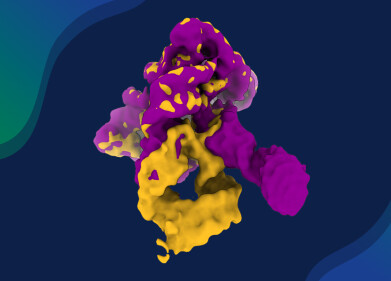
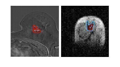 Side by side image of same breast tissue in MRI and FCI. (l) MRI image of breast with cancerous tumours circled in red (r) FCI image of same breast shows same tumour in red with secondary tumour spread in blue. Spread not visible in MRI. The patient had a mixed tumour i.e two different types of tumour and one of them is not visible in MRI. Photo Credit University of Aberdeen
 -
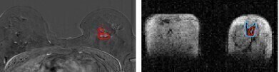 Photo Credit University of Aberdeen
 -
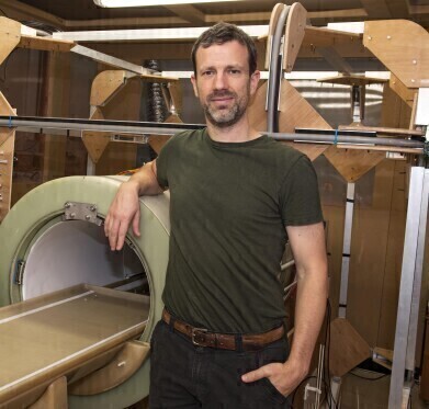 Dr Lionel Broche with the prototype FCI scanner. Photo Credit University of Aberdeen
Research news
Breakthrough scanner detects hidden cancer spread
Feb 26 2025
A pioneering scanner developed by scientists at the University of Aberdeen could transform breast cancer diagnosis and treatment, potentially reducing the need for repeat surgeries and enabling more personalised care.
Researchers from the University, in collaboration with NHS Grampian, used a prototype Field Cycling Imager (FCI) to examine breast tissue from newly diagnosed cancer patients. The FCI scanner demonstrated a higher accuracy in distinguishing tumour material from healthy tissue compared to conventional MRI methods.
This breakthrough has the potential to improve treatment outcomes for millions of patients. Currently, around 15% of women require a second surgery after a lumpectomy due to residual tumour cells. By providing more precise tumour mapping, FCI could help reduce the need for additional operations.
The success of FCI in breast cancer detection follows earlier promising results in identifying brain damage caused by stroke. Developed at the University of Aberdeen, FCI builds on MRI technology but operates at ultra-low magnetic fields, allowing it to detect disease-related changes in organs that were previously impossible to see.
Unlike conventional MRI, which uses strong magnetic fields to generate images, FCI can dynamically adjust magnetic field strength during a scan. This unique capability allows it to extract multiple layers of information from tissue, effectively functioning as multiple scanners in one. Additionally, FCI can detect tumours without the need for contrast agents, which are associated with potential kidney damage and allergic reactions in some patients.
Dr Lionel Broche, Senior Research Fellow in Biomedical Physics and lead researcher, highlighted the significance of these findings: “We found that FCI generates images that characterise breast tumours with greater accuracy. This could enhance biopsy precision, improve treatment planning, and reduce the need for repeat surgeries - offering significant benefits for patients.”
He added: “The University of Aberdeen pioneered the world’s first clinical MRI scanner in the 1970s, and it’s exciting to continue that legacy with an entirely new approach to imaging. As we refine FCI technology, its potential clinical applications are vast.”
Dr Gerald Lip, Consultant Radiologist at NHS Grampian and co-investigator in the study, recently appointed President of the British Society of Breast Radiology, commented: “These early results are promising, and further studies will help validate clinical applications. NHS Grampian treats 400 to 500 women with breast cancer annually, and the potential for FCI to reduce the need for additional surgeries could greatly benefit patients while improving resource efficiency.”
More information online
Published in Nature Communications Medicine


Digital Edition
Lab Asia Dec 2025
December 2025
Chromatography Articles- Cutting-edge sample preparation tools help laboratories to stay ahead of the curveMass Spectrometry & Spectroscopy Articles- Unlocking the complexity of metabolomics: Pushi...
View all digital editions


Events
Jan 21 2026 Tokyo, Japan
Jan 28 2026 Tokyo, Japan
Jan 29 2026 New Delhi, India
Feb 07 2026 Boston, MA, USA
Asia Pharma Expo/Asia Lab Expo
Feb 12 2026 Dhaka, Bangladesh


