Microscopy & microtechniques
A recent project conducted by in-house researchers at the UK’s national synchrotron facility on its Hard X-ray Nanoprobe beamline, looked at how microplastics waste may interact with zinc oxide (ZnO) nanomaterials in both freshwater and seawater scenarios. Their work published in Global Challenges revealed worrying environmental implications for aquatic organisms in the food chain.
Diamond accelerates electrons to produce intense light across the spectrum from UV and IR to X-rays that are used across instruments known as beamlines. Diamond has 34 beamlines grouped into eight science areas: macromolecular crystallography; soft condensed matter; cryo-biological imaging; imaging and microscopy; spectroscopy; crystallography; magnetic materials; structures and surfaces, enabling scientists to investigate the structure and properties of samples using a variety of imaging, scattering, diffraction and spectroscopic techniques.
The Imaging and Microscopy Group is made up of seven beamlines as well as the advanced Transmission Electron Microscopes (TEM) in the electron Physical Science Imaging Centre [ePSIC], which use electrons and X-rays to image samples under different experimental conditions across a range of length and time scales. The ability to image sample properties from atomic to macro scale using different modalities to extract information on phase, microstructure , chemical distributions, and under in situ conditions lends itself to a wide range of scientific areas, from chemistry and catalysis to environmental science, materials science, biology, medicine, and cultural heritage.
The research team used the Hard X-ray Nanoprobe beamline (Beamline I14) to look at how microplastics waste may interact with zinc oxide (ZnO) nanomaterials in freshwater and seawater scenarios. To make their study more relevant to the real world, the team tested a sunscreen containing zinc oxide, commonly used to block UV-radiation and a facial scrub containing tiny plastic beads – microbeads.
Their results confirmed that mixtures of Zn-aggregates and micro polymers were naturally leached and released from the commercial products revealing the potential for serious environmental implications. Fish and other aquatic organisms in the food chain can potentially ingest microplastics as well as zinc particles.
Lead author and beamline scientist, Miguel Gomez Gonzalez, explained that the team’s research revealed valuable information about how zinc oxide behaves in the environment and the dangers this poses to the food chain.
Explaining the impetus for the research, Miguel said that they had all seen how in recent decades, there has been a dramatic increase in the manufacture of engineered nanomaterials (particles about 1,000 times smaller than the diameter of a human hair), which has inevitably led to their dispersal into the environment. Zinc oxide (ZnO) is among one of the more abundant nanomaterials fabricated due to its use in electronics, semiconductors, and for antibacterial purposes. At the same time, plastic waste has become ubiquitous and may break down into smaller pieces called microplastics. These also are tiny, but ~100 times bigger than the nanomaterials.
Because both these elements are becoming more widespread in the environment, it is important to study what happens when they are potentially being combined in freshwater and seawater, in order to make environmental risk assessments more accurate.
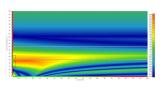
Figure 2: X-ray fluorescence (XRF, left) maps (100 nm pixel size) for the Zn signal and differential phase contrast (DPC, right) image for morphological inspection of the adsorbed structures measured at the hard X-ray nanoprobe (I14 beamline). Zn-particles from the sunscreen were deposited on the pristine microplastics after incubation in seawater (top row) while ZnO nanomaterials were deposited on the microplastics leached from the exfoliating cleanser after incubation in seawater as well (bottom row).
Images are an adaptation of the published paper at:
https://onlinelibrary.wiley.com/doi/full/10.1002/gch2.202300036.
The team took some pure zinc oxide particles (ranging from 80 to 200 nm size) and incubated them in different kinds of environmental solutions for a week, allowing their natural stabilisation. They then mixed them with small polystyrene microspheres (~900 mm diameter, about the size of a grain of sand), or the microbeads from the facial scrub, and stirred them together for a day. The sunscreen was also incubated in the environmental solutions for a week and then added the microplastics for a day. The objective was to check if the zinc oxide could leach out of the sunscreen and adhere to the microplastics.
After washing and rinsing the microplastics, it was found that the zinc oxide was adsorbed to the plastic surfaces. This was observed by scanning electron microscopy (SEM), (Figure 1). This confirmed that microplastics and zinc oxide can interact in our water bodies, which might affect how they impact the environment.
The team then examined these zinc oxide covered microplastics at the Hard X-ray Nanoprobe beamline. Here X-rays are focused to a nanometre scale beam (50nm) and a technique called nano- X-ray Fluorescence (n-XRF) is used to build a map of different elements as a sample is scanned through the beam. This was used to create a nanometre scale map of the distribution of the Zinc on the surface of the microplastics (Figure 2). An advantage of using the nanoprobe beamline is the ability to combine information from different techniques, here the n-XRF could be combined with differential phase contrast imaging to show the morphology of the adsorbed nanomaterials
A further X-ray technique called X-ray absorption near-edge structure spectroscopy (XANES) was applied at I14 to investigate spatially resolved chemical changes and transformations that may have occurred to the zinc oxide following adsorption to the microplastics and incubation in a freshwater solution.
Miguel concluded,: “We found that the zinc oxide had transformed into different types of zinc-related particles. Some of these new particles (Zn-sulphide) were formed quickly, while others formed more slowly but were more stable (Zn-phosphate) (Figure 3). This reveals valuable information about how zinc oxide behaves when it is in the environment.”
Miguel commented: “The ability of zinc oxide, both pure nanomaterials and those released from a sunscreen, to stick to very small pieces of plastic has big implications. These plastics can even come from everyday items like exfoliating facial cleansers. In this study, we found the microplastics can carry even smaller particles of zinc from place to place. As a consequence, fish or other aquatic organisms could swallow these microplastics, ingesting zinc particles at the same time.
“We need to understand how this engineered zinc oxide changes when it gets into freshwaters and how much of it can stick to small plastic wastes. This is important for making everyone aware, from people who make these products to those who regulate them, about the potential harm they could do to our environment. Better rules for managing waste are needed, especially related to tiny particles like these. As we continue to produce more and more of these micro- and nanoparticles, their effect on our environment is going to keep growing. Because they are so long-lasting, they can pose a risk to different organisms, and ultimately even make their way into our food. This is something we simply cannot afford to ignore.”
The Diamond team included a student, Tatiana Da-Silva Ferreira, who at the time of research was attending Edinburgh University and was part of Diamond’s 12- week Summer Placement scheme. The popular programme allows undergraduate students studying for a degree in science, engineering, computing or mathematics to gain experience working in a number of different teams at Diamond.
Miguel praised Tatiana, who is now studying for a PhD in Switzerland, for her key contribution to the start of this environmental project. “Tatiana did a great job in optimising the conditions for the seven days stabilisation of nanomaterials, followed by the 24-hours incubation of microplastics and nanomaterials. In addition, she improved the filtering protocol and isolation of the microplastics after the incubation period. Likewise, she performed the very preliminary scanning electron microscopy analysis which revealed nanomaterials adsorption into the plastic surfaces. Therefore, her contribution was key for the overall success of this environmentally relevant project.”
Diamond Light source allows users to access many different imaging techniques and modalities. Eight different facilities can be used to study samples, with various sizes and compositions, from large engineering components down to tiny cells and nanoparticles. Imaging techniques available at Diamond exploit the penetrating power of X-rays, allowing you to probe both interior structure of materials, and see internal or hidden components. Using different X-ray and Electron imaging techniques information can be obtained, at micro to atomic resolutions, of structure and porosity, crystal phase, chemical composition and speciation.
Related publication: Gomez-Gonzalez, M. A., Da Silva-Ferreira, T., Clark, N., Clough, R., Quinn, P. D., Parker, J. E., Toward Understanding the Environmental Risks of Combined Microplastics/Nanomaterials Exposures: Unveiling ZnO Transformations after Adsorption onto Polystyrene Microplastics in Environmental Solutions.
Global Challenges 2023, 2300036.
https://doi.org/10.1002/gch2.202300036
1. I12 The Joint Engineering, Environmental and Processing (JEEP) beamline (I12) produces high energy X-rays (53-150 keV) for imaging and diffraction of large or dense samples, and in situ processes such as high temperature or applying stress and strain that require large experimental rigs to be installed. I12 enables large complex experiments across fields including material, life, and heritage sciences.
2. I13-2 Imaging beamline is focussed on micro-and nano-tomography and provides multiscale imaging capabilities over three orders of magnitude in resolution. The beamline operates in the 8-30 keV energy range, offering a variety of imaging capabilities including in-line phase contrast imaging, full-field microscopy and grating interferometry.
3. 113-1 Coherence beamline is a hard X-ray beamline, operating in the 6-20 keV range. It specialises in high-speed, multiscale and multimodal coherent diffraction imaging and ptychography in both transmission and Bragg geometries. These methods provide nanoscale resolutions with a quantitative phase contrast and can often be combined with methods, such as tomography and XANES, and complementary signals, such as fluorescence and diffraction mapping.
4. I08 Scanning X-ray Microscopy beamline is for morphological, elemental and chemical speciation on a broad range of organic-inorganic interactions in a 250 - 4400 eV photon energy range, and sample investigations under ambient conditions. I08 has a range of applications including biological and biomedical sciences, earth and environmental science, geochemistry, and materials science.
5. I08-1 Soft X-ray spectro- and tomo-ptychography branchline (operating in the 250-2000 eV photon energy range) is a recent facility, growing in capabilities. Key developments focus on multi-dimensional spectroscopic ptychographic imaging of light elements including variable polarisation of X-rays.
6. I14, the Hard X-ray Nanoprobe beamline, provides a focussed beam of 50 nm for high resolution imaging of a wide range of samples using multiple techniques including X-ray fluorescence, diffraction, X-ray Absorption Near Edge Structure (XANES), differential phase contrast imaging and ptychography. The beamline has a strong focus on in situ characterisation and provides environments for gas flow, heating, liquid and electrochemistry, while the flexible end station space enables integration of customised user sample environments.
7. DIAD (Dual Imaging and Diffraction) beamline provides simultaneous imaging and diffraction capabilities for advanced material and biological research. The beamline features a unique dual-beam setup, where one beam is optimised for high-resolution imaging, allowing for detailed visualisation of internal features and defects, while the other beam is tailored for diffraction, providing insights into the crystalline structure and phase composition. It supports both static and dynamic studies, enabling time-resolved experiments that can capture the evolution of materials under stress, temperature changes, and other environmental factors.
8. Electron Physical Science Imaging Centre (ePSIC) is a national facility for aberration corrected electron microscopy, established as a collaboration between Diamond, the University of Oxford and Johnson Matthey. ePSIC is equipped with two aberration corrected microscopes for studying materials at the atomic scale. The JEOL ARM300F is a double-corrected microscope dedicated to atomic scale imaging and 4D STEM studies. The JEOL ARM200F is a probe-corrected microscope that specializes in spectroscopic imaging with both EELS and EDX spectrometers.
For further information about Diamond’s Imaging and Microscopy group please visit:
https://www.diamond.ac.uk/Instruments/Imaging-and-Microscopy.html or contact Julia Parker, Science Group leader.


.jpg)
-(2).jpg)


.jpg)