Microscopy & microtechniques
Report on the Use of AFM and Single-Cell Force Spectroscopy
Jan 23 2013
The interdisciplinary Nanoscience Center (iNANO) was formed by various research groups at Aarhus University together with groups from the Faculty of Science at Aalborg University. iNANO comprises facilities for the synthesis of nanostructured and nanopatterned 0D (i.e. nanoparticle), 1D, 2D and 3D materials.
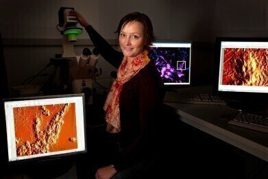
The group of Dr Rikke Meyer works at the interface between microbiology and nanoscience in the quest to understand how bacteria form biofilms and how this may be prevented. AFM and optical microscopy are used to visualise bacterial cells and to study the interaction forces between cells and an abiotic substrate.
The motivation for using AFM in Dr Meyer's research was firstly to obtain detailed images of bacterial cells without extensive sample preparation. Furthermore, as she is interested in the interactions between bacteria and abiotic surfaces, she and her team use AFM force spectroscopy to quantify these interaction forces. AFM is one of several techniques used in these studies. These also include brightfield microscopy, fluorescence microscopy, confocal laser scanning microscopy, scanning electron microscopy and transmission electron microscopy.
Dr Meyer commented on her research and reasons behind her choice of AFM: "The coupling with optical microscopy is no doubt the feature that was most important for me in deciding to go with an AFM from JPK. As a microbiologist, I work with very heterogenous samples and it is not feasible to use AFM imaging to locate the field of interest, as large areas of the sample are often visualised to locate a site of interest. In the combined system, we can use the optical image to locate cells of interest before engaging the AFM for imaging or other measurements."
She continued: "AFM has mostly been used to study bacterial cells that are isolated in pure culture. However, the vast majority of the bacterial species we know to date have not been isolated and can only be studied in situ. Fluorescence labelling allows a rough identification of bacteria directly in the sample and fluorescence imaging can thus be used to locate cells of interest before AFM imaging begins. The combination of AFM with optical imaging is thus particularly important for the analysis of bacteria in environmental samples."
.jpg)
-(2).jpg)

Digital Edition
Lab Asia Dec 2025
December 2025
Chromatography Articles- Cutting-edge sample preparation tools help laboratories to stay ahead of the curveMass Spectrometry & Spectroscopy Articles- Unlocking the complexity of metabolomics: Pushi...
View all digital editions


Events
Jan 21 2026 Tokyo, Japan
Jan 28 2026 Tokyo, Japan
Jan 29 2026 New Delhi, India
Feb 07 2026 Boston, MA, USA
Asia Pharma Expo/Asia Lab Expo
Feb 12 2026 Dhaka, Bangladesh


