-
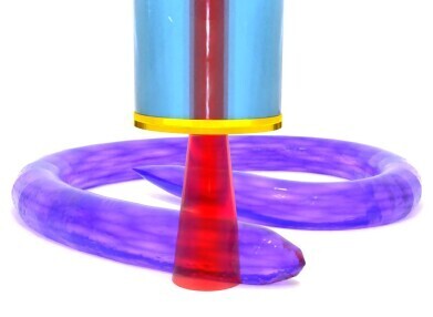 Caenorhabditis elegans (Credit: University of Nottingham)
Research News
Microscopic probe holds promise for early cancer diagnosis
May 23 2024
Researchers at the University of Nottingham have created a world-first endoscopic device with the ability to detect changes in cell hardness, an indication which could suggest the presence of tumour cells.
Softer than normal cells, early stage cancer cells are able to squeeze through tight gaps and rapidly spread throughout the body, known as metastasis. During this process, collections of cells modify their surrounding environment to create stiff tumours that protect them from outside threats.
The new device can 3Dimage individual cells with a hair-thin endoscopic probe, meaning it will be possible to perform histology (i.e. investigating microscopic cellular tissue) based on abnormal stiffness at the single cell level inside the human body for the first time.
Dr Salvatore La Cavera III, lead-author and Nottingham Research Fellow in the Optics and Photonics Group at the University of Nottingham, said: “We aim to develop new endoscopic technologies that make diagnostics faster, safer and clearer for both patients and clinicians. Typically, histopathology requires destructive, invasive biopsies that are not only uncomfortable and potentially damaging for the patient, but require significant logistics such as chemical processing, transportation and analysis.
“Our device makes it possible to ‘feel for a stiff lump,’ but on a single cellular scale, meaning we could catch cancer early at microscopic cell scales rather than large malignant tumour scale. It is non-invasive, non-toxic and very promisingly, is related to technology that can quantitively determine the presence of cancer cells using artificial intelligence – providing a chronically understaffed area with a much-needed solution to a real-world problem that the industry has faced for decades.”
The tool achieves an high imaging resolution, detecting the stiffness of objects down to billionths of a metre (nanometres), through a physical phenomenon called Brillouin scattering, where a laser beam interacts with the natural stiffness of a specimen. The team used this to help biologists visualise the 3D stiffness of a microscopic organism, called Caenorhabditis elegans - a free-living worm, known scientifically as a nematode. They were able to provide visualisation and material information about an elusive part of the organism’s anatomy, the cuticle, that up until now had only been imaged through electron microscopes in non-living conditions.
Dr Veeren Chauhan co-author and Assistant Professor in Whole Organism Analytics at the School of Pharmacy at the University of Nottingham, said: “We have demonstrated the capabilities of this exciting next-generation technology to reveal the physical surface properties of a fully formed microscopic organism in unprecedented detail. Nematodes are an excellent model for human biology and are considered to be the most completely understood animal on the planet in terms of genetics, neurology and developmental biology.”
The new probe, she added, had the potential for enabling researchers to follow the development of an individual nematode's physical surface properties throughout its entire life cycle, from egg to adult, in approximately three days, which would take upwards of 18 years when considering human development.
More information online


.jpg)
Digital Edition
Lab Asia 31.6 Dec 2024
December 2024
Chromatography Articles - Sustainable chromatography: Embracing software for greener methods Mass Spectrometry & Spectroscopy Articles - Solving industry challenges for phosphorus containi...
View all digital editions


Events
Jan 22 2025 Tokyo, Japan
Jan 22 2025 Birmingham, UK
Jan 25 2025 San Diego, CA, USA
Jan 27 2025 Dubai, UAE
Jan 29 2025 Tokyo, Japan


