Microscopy & Microtechniques
Launch of New Cryo-Correlative Cooling Stage
Feb 07 2013
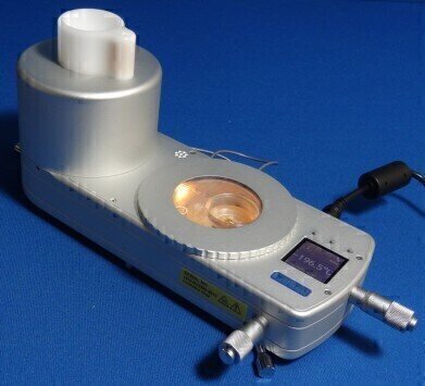
Linkam are pleased to announce the development and launch of a stage for cryo-correlative light/electron microscopy (cryo-CLEM). It was designed in collaboration with scientists at the Leiden University Medical Centre (LUMC) led by Bram Koster and Erik Bos. Cryo-CLEM is the correlation of images captured with a cryostage on a fluorescent light microscope and images of the same sample observed with Transmission Electron Microscopy (TEM).
Dr Lucy Collinson of the London Research Institute (LRI) and Cancer Research UK (CRUK), is using the correlative stage as part of a workflow that starts with cells expressing cancer-related genes and ends with imaging in synchrotrons located in Oxford, Berlin, and Barcelona. Within the synchrotrons, the scientists are able to image the cells using a cutting-edge technique called 'soft X-ray tomography'. The LRI focuses on three themes of research: the biology of tissues and tumours, cellular regulatory mechanisms and genomic integrity & cell cycle.
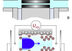
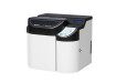
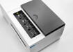
The correlative stage can hold samples at a stable -196°C enabling scientists to study TEM grid samples at 100x magnification, identifying areas of further interest, and facilitating the movement of these analysed grids to the TEM. With automated liquid nitrogen control, heated optics and a digital display the unit is a compact and efficient system for this work.
In particular, Dr Collinson is working with Dr Sharon Tooze, Head of the Secretory Pathways Laboratory at the LRI, to study cells that are undergoing autophagy, which is a normal process involving degradation and recycling of unnecessary or dysfunctional cell components
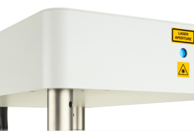
With Dr Liz Duke, a Principal Beamline Scientist at the Diamond Light Source synchrotron in Oxfordshire, they have developed a process for imaging these fluorescent proteins in cells as close to their living state as possible, considering that they must place them in a vacuum to take very high magnification images. They grow the cells on very thin carbon films attached to a 3mm diameter gold grid and freeze the cells in liquid ethane at –174°C.
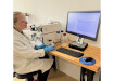
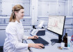
The correlative stage is used to image the fluorescence in the cells while they are still frozen. The frozen grids are then shipped to the synchrotron, where they put them into the soft X-ray microscope so that they can image the structure of the entire cell in 3D.
Digital Edition
International Labmate Buyers' Guide 2024/25
June 2024
Buyers' Guide featuring: Product Listings & Manufacturers Directory Chromatography Articles - Enhancing HPLC Field Service with fast-response, non-invasive flowmeters - Digital transformatio...
View all digital editions


Events
Jul 03 2024 Gandhinagar, India
Jul 07 2024 Dublin, Ireland
Jul 20 2024 Denver, CO, USA
Jul 21 2024 Cape Town, South Africa
Jul 28 2024 San Diego, CA USA


